应用化学 ›› 2025, Vol. 42 ›› Issue (8): 1035-1056.DOI: 10.19894/j.issn.1000-0518.250087
• 综合评述 • 下一篇
黄欣欣1, 石有圣2, 邓涛3, 蔡春1( )
)
收稿日期:2025-03-01
接受日期:2025-06-11
出版日期:2025-08-01
发布日期:2025-08-11
通讯作者:
蔡春
Xin-Xin HUANG1, You-Sheng SHI2, Tao DENG3, Chun CAI1()
Received:2025-03-01
Accepted:2025-06-11
Published:2025-08-01
Online:2025-08-11
Contact:
Chun CAI
About author:c.cai@njust.edu.cn摘要:
聚集诱导发光(AIE)受到了越来越多的关注,并已被广泛应用于生化分析、光电材料等多个领域。 不同于传统的荧光探针,AIE型荧光探针聚集后,分子内运动由于空间位阻而受限,从而表现出强发光。AIE型荧光探针表现出更高的选择性、灵敏度和信噪比,以及更短的响应时间。 这些优势使得聚集诱导发光广泛应用于生化检测、疾病的早期诊疗和细胞成像等多个方面。 本文总结了AIE型荧光探针在酶、金属离子和生物活性小分子等生物标志物检测中的应用。 此外,还讨论了AIE型荧光探针面临的挑战和未来的发展前景。
中图分类号:
黄欣欣, 石有圣, 邓涛, 蔡春. 聚集诱导发光型荧光探针在生物标志物检测中的研究进展[J]. 应用化学, 2025, 42(8): 1035-1056.
Xin-Xin HUANG, You-Sheng SHI, Tao DENG, Chun CAI. Research Progress of Fluorescent Probes Based on Aggregation-Induced Emission for Detection of Biomarkers[J]. Chinese Journal of Applied Chemistry, 2025, 42(8): 1035-1056.
| Diseases | Biomarkers |
|---|---|
| Lung cancer | Carcinoembryonic antigen (CEA), neuron specific enolase (NSE), squamous cell carcinoma antigen (SCCA), cytokeratin 19 fragment antigen (CYFRA21-1), biothiols etc. |
| Liver cancer | Alpha fetoprotein (AFP), alpha-L fucosidase (AFU), hepatocyte growth factor (HGF) etc. |
| Gastric cancer | Carcinoembryonic antigen (CEA), carbohydrate antigen (CA 19-9, CA72-4), pepsinogen Ⅰ/Ⅱ(PG Ⅰ/Ⅱ) etc. |
| Breast cancer | Carcinoembryonic antigen (CEA), carbohydrate antigen (CA), tissue polypeptide specific antigen (TPS), epithelial cadherin (E-cadherin), alkaline phosphatase (ALP), nitroreductase (NTR), H2S etc. |
| Cervical cancer | Carbohydrate antigen (CA125,CA72-4), squamous cell carcinoma antigen (SCCA), hepatocyte growth factor (HGF), mixed lineage kinase domain-like protein (MLKL), Cu2+, Hg2+etc. |
| Ovary cancer | Tumor necrosis factor-α (TNF-α), humanepididymisprotein4 (HE4), hexokinase2 (HK2), sequestosome-1 (SQSTM1/p62),β-galactosidase (β-Gal) etc. |
| Colorectal cancer | Carcinoembryonic antigen (CEA), lactate dehydrogenase (LDH), microtubule-associated protein 1 light chain 3 beta (MAP1LC3B), unc-51 like autophagy activating kinase 1 (ULK-1), glutathione (GSH) etc. |
| Cardiovascular disease | Myoglobin (Myo), cardiac troponin (cTn), creatine kinase isoenzyme (CK-MB), B-type natriuretic peptide (BNP), growth differentiation factor 15 (GDF-15) etc. |
| Alzheimer's disease | Amyloid-β peptides (Aβ peptides), tau protein, apolipoprotein E (apoE), circulating free microRNAs (miRNAs), alpha-1 antitrypsin (AAT), Zn2+etc. |
| Inflammation | C-reactive protein (CRP), serum amyloid A (SAA), procalcitonin (PCT), ferritin (SF), reactive oxygen species (ROS) etc. |
表1 目前公认的部分生物标志物及相关疾病
Table 1 Examples of universally acknowledged biomarkers and related diseases
| Diseases | Biomarkers |
|---|---|
| Lung cancer | Carcinoembryonic antigen (CEA), neuron specific enolase (NSE), squamous cell carcinoma antigen (SCCA), cytokeratin 19 fragment antigen (CYFRA21-1), biothiols etc. |
| Liver cancer | Alpha fetoprotein (AFP), alpha-L fucosidase (AFU), hepatocyte growth factor (HGF) etc. |
| Gastric cancer | Carcinoembryonic antigen (CEA), carbohydrate antigen (CA 19-9, CA72-4), pepsinogen Ⅰ/Ⅱ(PG Ⅰ/Ⅱ) etc. |
| Breast cancer | Carcinoembryonic antigen (CEA), carbohydrate antigen (CA), tissue polypeptide specific antigen (TPS), epithelial cadherin (E-cadherin), alkaline phosphatase (ALP), nitroreductase (NTR), H2S etc. |
| Cervical cancer | Carbohydrate antigen (CA125,CA72-4), squamous cell carcinoma antigen (SCCA), hepatocyte growth factor (HGF), mixed lineage kinase domain-like protein (MLKL), Cu2+, Hg2+etc. |
| Ovary cancer | Tumor necrosis factor-α (TNF-α), humanepididymisprotein4 (HE4), hexokinase2 (HK2), sequestosome-1 (SQSTM1/p62),β-galactosidase (β-Gal) etc. |
| Colorectal cancer | Carcinoembryonic antigen (CEA), lactate dehydrogenase (LDH), microtubule-associated protein 1 light chain 3 beta (MAP1LC3B), unc-51 like autophagy activating kinase 1 (ULK-1), glutathione (GSH) etc. |
| Cardiovascular disease | Myoglobin (Myo), cardiac troponin (cTn), creatine kinase isoenzyme (CK-MB), B-type natriuretic peptide (BNP), growth differentiation factor 15 (GDF-15) etc. |
| Alzheimer's disease | Amyloid-β peptides (Aβ peptides), tau protein, apolipoprotein E (apoE), circulating free microRNAs (miRNAs), alpha-1 antitrypsin (AAT), Zn2+etc. |
| Inflammation | C-reactive protein (CRP), serum amyloid A (SAA), procalcitonin (PCT), ferritin (SF), reactive oxygen species (ROS) etc. |
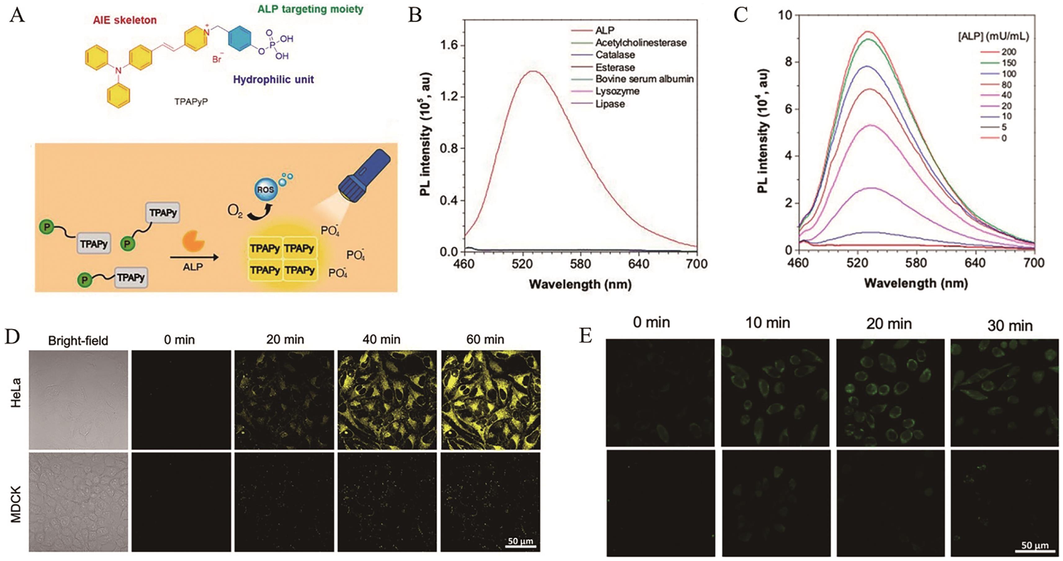
图1 (A) AIE探针TPAPyP的结构及其响应机理; (B)探针TPAPyP(100 μmol/L)与不同分析物孵育后的荧光响应; (C)探针TPAPyP(100 μmol/L)与不同浓度ALP共同孵育后的荧光光谱; (D)探针TPAPyP(2 μmol/L)在HeLa细胞和MDCK细胞中的荧光成像; (E) ROS探针DCFH在HeLa细胞(上)和MDCK细胞(下)中的荧光成像[34]
Fig.1 (A) The structure and response mechanism of AIE probe TPAPyP; (B) The fluorescence response of probe TPAPyP (100 μmol/L) after incubation with different analytes; (C) The fluorescence spectra of probe TPAPyP (100 μmol/L) after reaction with various concentrations of ALP; (D) The fluorescence imaging of HeLa cells and MDCK cells incubated with probe TPAPyP (2 μmol/L); (E) The fluorescence imaging of HeLa cells (top) and MDCK cells (bottom) incubated with ROS probe DCFH[34]
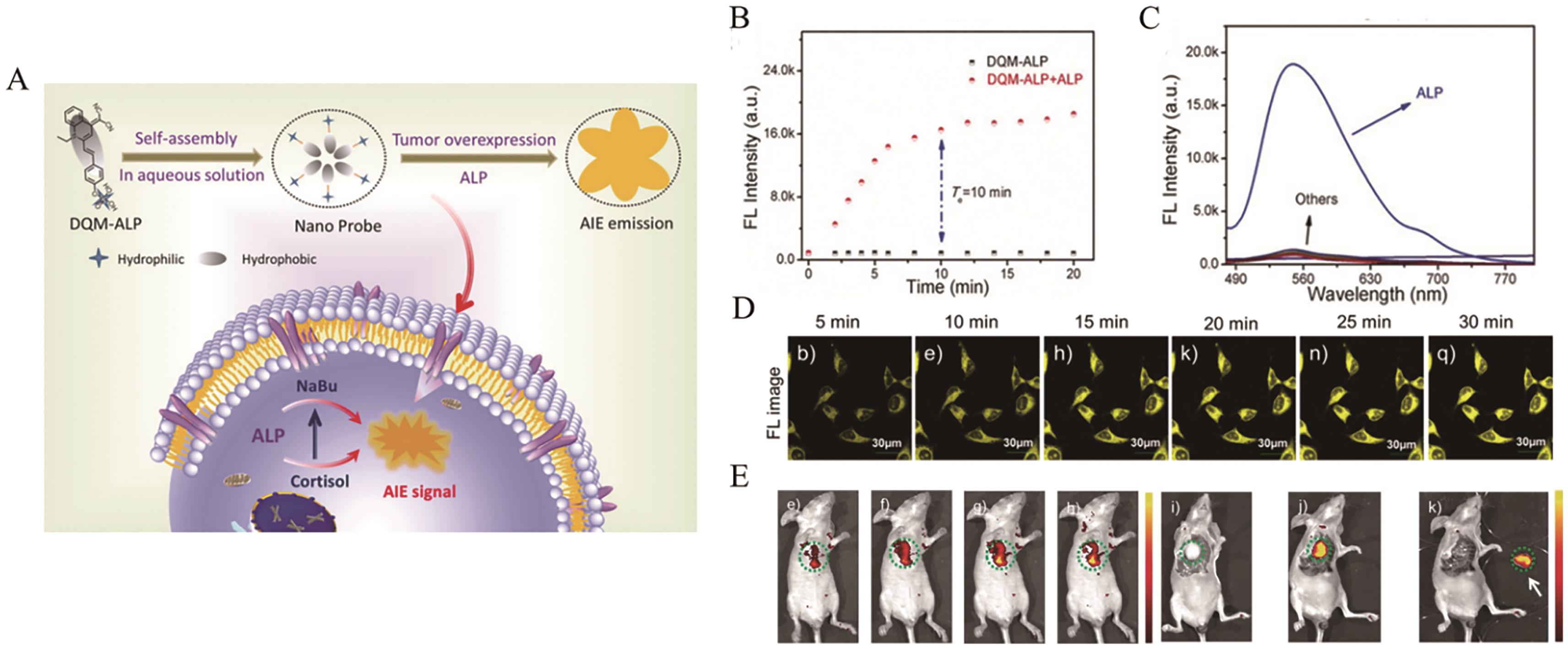
图2 (A) AIE探针DQM-ALP的结构及其在细胞内的响应示意图; (B)探针DQM-ALP与ALP响应前后的动力学曲线; (C)探针DQM-ALP(10 μmol/L)与不同分析物孵育后的荧光响应; (D)探针DQM-ALP(10 μmol/L)在HeLa细胞中的荧光成像; (E)探针DQM-ALP(50 μmol/L)在小鼠肝癌肿瘤组织中的荧光成像[35]
Fig.2 (A) The structure and schematic illustration of intracellular response of AIE probe DQM-ALP; (B) The kinetic curves of probe DQM-ALP before and after response to ALP; (C) The fluorescence response of probe DQM-ALP (10 μmol/L) after incubation with different analytes; (D) The fluorescence imaging of HeLa cells incubated with probe DQM-ALP (10 μmol/L); (E) Fluorescence imaging of liver tumor-bearing mice after injection of probe DQM-ALP (50 μmol/L)[35]
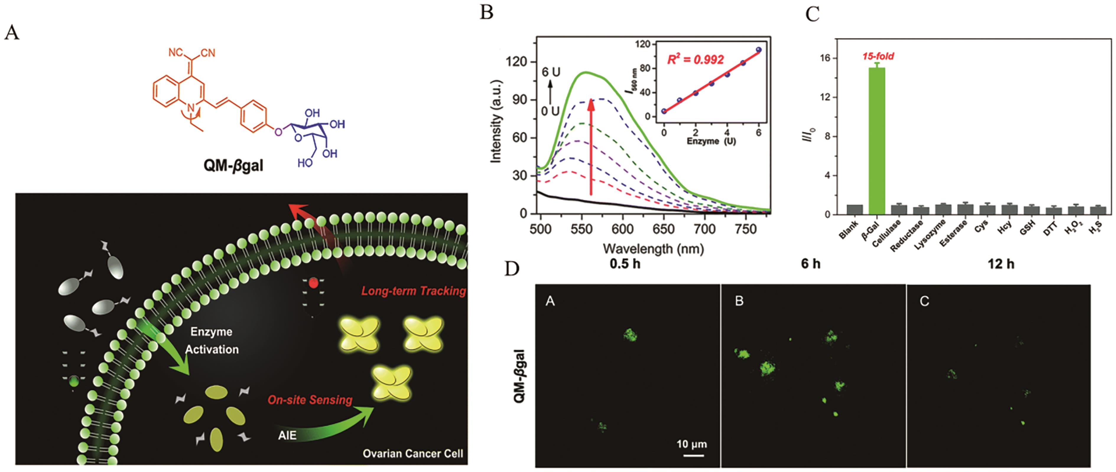
图3 (A) AIE探针QM-βgal的结构及其在细胞内的响应示意图; (B)探针QM-βgal(10 μmol/L)与不同浓度β-Gal响应后的荧光光谱及相应的线性关系; (C)探针QM-βgal(10 μmol/L)与不同分析物孵育后的荧光响应; (D)探针QM-βgal(10 μmol/L)在SKOV3细胞中的荧光成像[37]
Fig.3 (A) The structure and schematic illustration of intracellular response of AIE probe QM-βgal; (B) The fluorescence spectra of probe QM-βgal (10 μmol/L) after reaction with various concentrations of β-Gal and corresponding linear relationship; (C) The fluorescence response of probe QM-βgal (10 μmol/L) after incubation with different analytes; (D) The fluorescence imaging of SKOV-3 cells incubated with probe QM-βgal (10 μmol/L)[37]
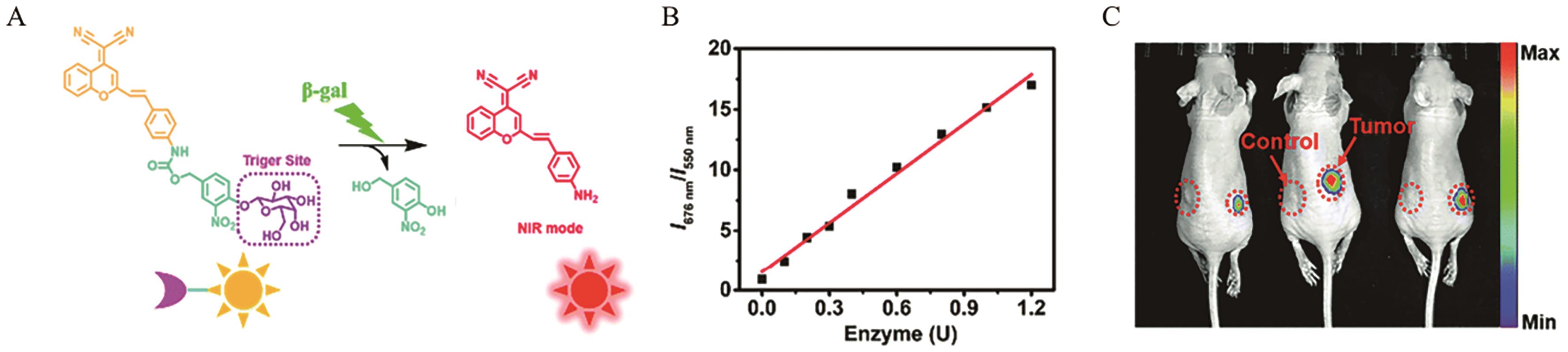
图4 (A) AIE探针DCMCA-β-gal的结构及其响应机理; (B)探针DCMCA-β-gal的比率荧光信号I676 nm/I550 nm与β-Gal浓度之间的线性关系; (C)探针DCMCA-β-gal在小鼠卵巢肿瘤中的荧光成像[38]
Fig.4 (A) The structure and response mechanism of AIE probe DCMCA-β-gal; (B) The linear relationship between the ratio-metric fluorescence intensity I676 nm/I550 nm of probe DCMCA-β-gal and the concentration of β-Gal; (C) Fluorescence imaging of ovarian tumor-bearing mice after injection of probe DCMCA-β-gal[38]
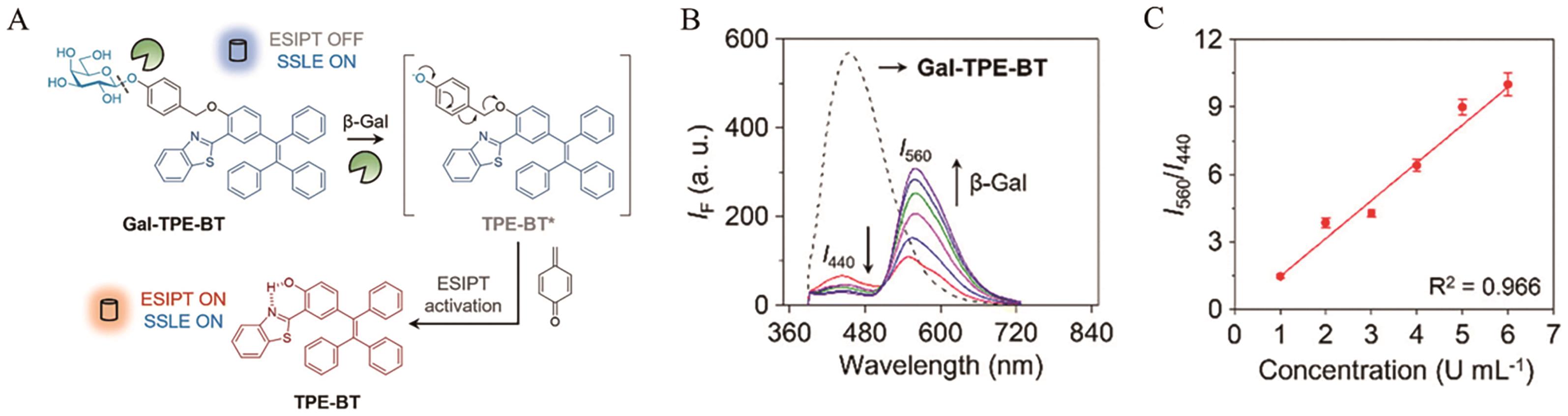
图5 (A) AIE探针Gal-TPE-BT的结构及其响应机理; (B)探针Gal-TPE-BT(10 μmol/L)与不同浓度β-Gal响应后的荧光光谱; (C)探针Gal-TPE-BT的比率式荧光信号I550 nm/I440 nm与β-Gal浓度之间的线性关系[39]
Fig.5 (A)The structure and response mechanism of AIE probe Gal-TPE-BT; (B) The fluorescence spectra of probe Gal-TPE-BT (10 μmol/L) after reaction with various concentrations of β-Gal; (C) The linear relationship between the ratio-metric fluorescence intensity I550 nm/I440 nm of probe Gal-TPE-BT and the concentration of β-Gal[39]
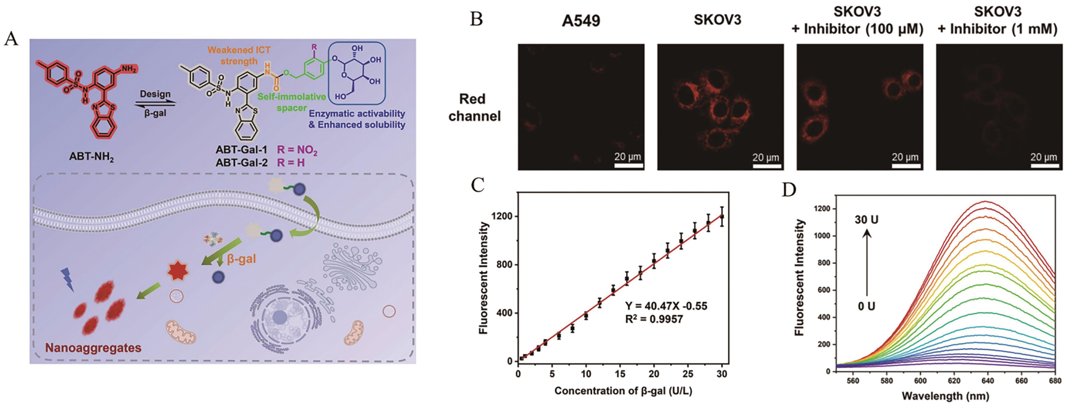
图6 (A) AIE探针ABT-Gal的结构及其在细胞内的响应示意图; (B)探针ABT-Gal-1(20 μmol/L)在A549细胞和SKOV-3细胞中的荧光成像; (C)探针ABT-Gal-1的荧光强度与β-Gal浓度之间的线性关系; (D)探针ABT-Gal-1(10 μmol/L)与不同浓度β-Gal响应后的荧光光谱[40]
Fig.6 (A) The structure and schematic illustration of intracellular response of AIE probe ABT-Gal; (B) The fluorescence imaging of A549 cells and SKOV-3 cells incubated with probe ABT-Gal-1 (20 μmol/L); (C) The linear relationship between the fluorescence intensity of probe ABT-Gal-1 and the concentration of β-Gal; (D) The fluorescence spectra of probe ABT-Gal-1 (10 μmol/L) after reaction with various concentrations of β-Gal[40]
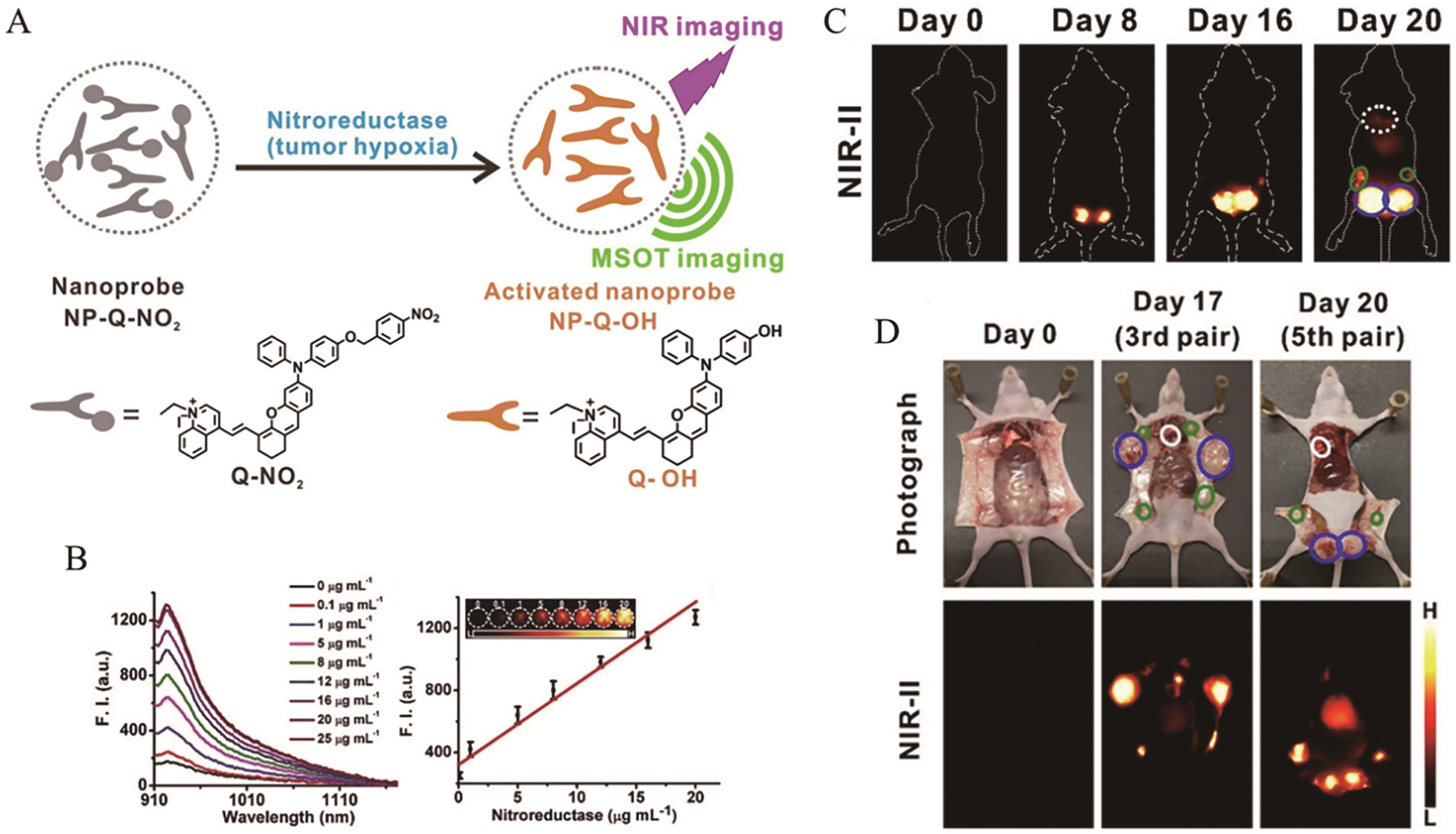
图7 (A) AIE探针Q-NO2的结构及其响应机理; (B)探针Q-NO2(10 μmol/L)与不同浓度硝基还原酶响应后的荧光光谱及相应的线性关系; (C)探针Q-NO2在小鼠乳腺肿瘤中的荧光成像; (D)探针Q-NO2在小鼠肿瘤中的荧光成像[41]
Fig.7 (A) The structure and response mechanism of AIE probe Q-NO2; (B) The fluorescence spectra of probe Q-NO2 (10 μmol/L) after reaction with various concentrations of NTR and corresponding linear relationship; (C) Fluorescence imaging of breast tumor-bearing mice after injection of probe Q-NO2; (D) Fluorescence imaging of tumor-bearing mice after injection of probe Q-NO2[41]
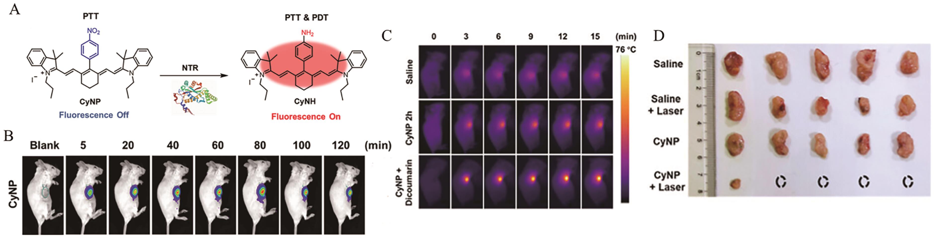
图8 (A) AIE探针CyNP的结构及其响应机理; (B)探针CyNP在小鼠乳腺肿瘤中的荧光成像; (C)探针CyNP在小鼠乳腺肿瘤中的光热成像; (D)小鼠中肿瘤的图像[42]
Fig.8 (A) The structure and response mechanism of AIE probe CyNP; (B) Fluorescence imaging of breast tumor-bearing mice after injection of probe CyNP; (C) Photothermal imaging of breast tumor-bearing mice after injection of probe CyNP; (D) Photos of tumor in mice[42]
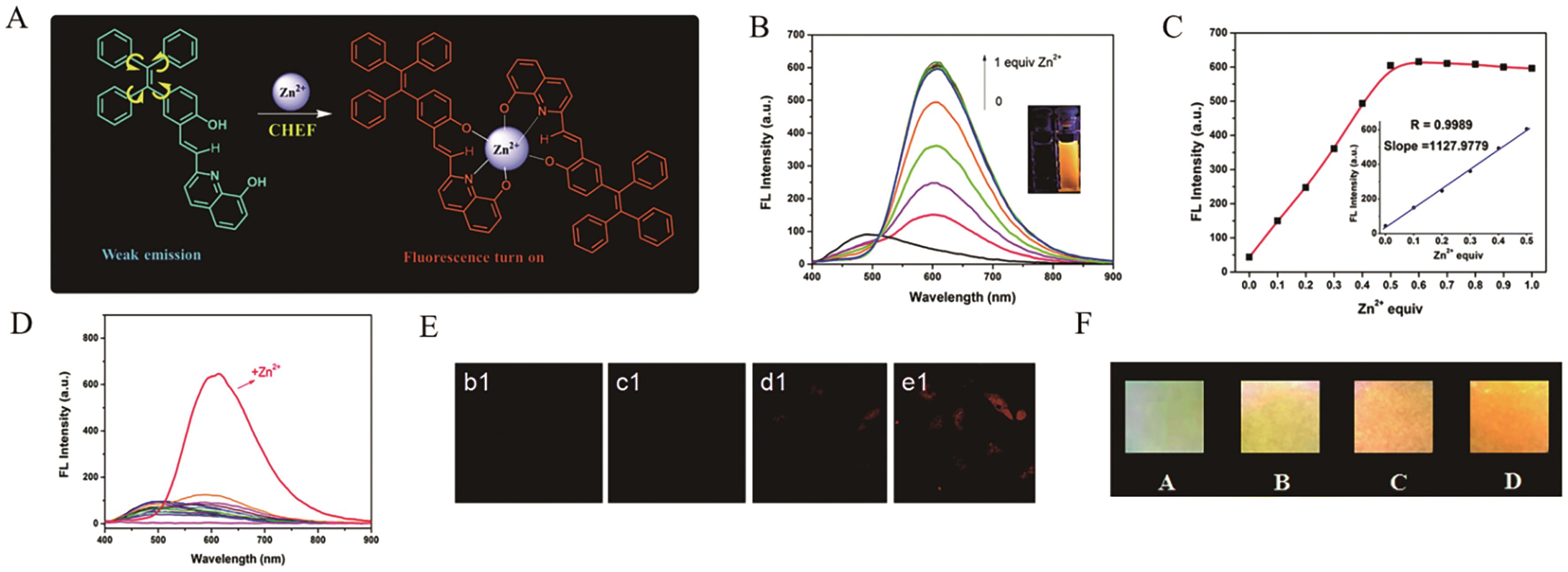
图9 (A) AIE探针TPE(OH)-8HQ的结构及其响应机理; (B)探针TPE(OH)-8HQ(10 μmol/L)与不同浓度Zn2+响应后的荧光光谱; (C)探针TPE(OH)-8HQ的荧光强度与Zn2+浓度之间的线性关系; (D)探针TPE(OH)-8HQ(10 μmol/L)与不同分析物孵育后的荧光响应; (E)探针TPE(OH)-8HQ(20 μmol/L)在HeLa细胞中的荧光成像; (F)探针TPE(OH)-8HQ(1 mmol/L)在试纸上对Zn2+的荧光响应图像[47]。
Fig.9 (A) The structure and response mechanism of AIE probe TPE(OH)-8HQ; (B) The fluorescence spectra of probe TPE(OH)-8HQ (10 μmol/L) after reaction with various concentrations of Zn2+; (C) The linear relationship between the fluorescence intensity of probe TPE(OH)-8HQ and the concentration of Zn2+; (D) The fluorescence response of probe TPE(OH)-8HQ (10 μmol/L) after incubation with different analytes; (E) The fluorescence imaging of HeLa cells incubated with probe TPE(OH)-8HQ (20 μmol/L); (F) Fluorescence response of TPE(OH)-8HQ (1 mmol/L) to Zn2+ on test papers[47]
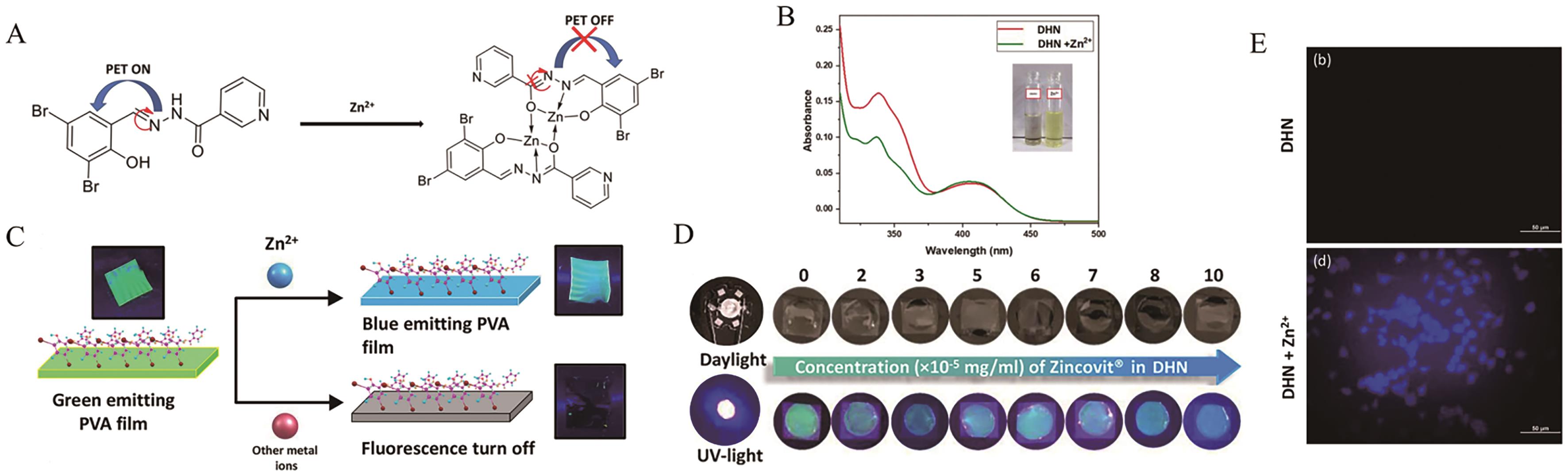
图10 (A) AIE探针DHN的结构及其响应机理; (B)探针DHN(10 μmol/L)与Zn2+响应前后的吸收光谱; (C) PVA_DHN复合薄膜的检测机理; (D)在紫外照射下DHN响应不同浓度Zincovit的照片; (E)探针DHN(5 μmol/L)在N2a细胞中的荧光成像[48]
Fig.10 (A) The structure and response mechanism of AIE probe DHN; (B) The absorption spectra of probe DHN(10 μmol/L) before and after response to Zn2+; (C) The detection mechanism of PVA_DHN composite film; (D) Photos of DHN response to various concentrations of Zincovit under UV irradiation; (E) The fluorescence imaging of N2a cells incubated with probe DHN (5 μmol/L)[48]
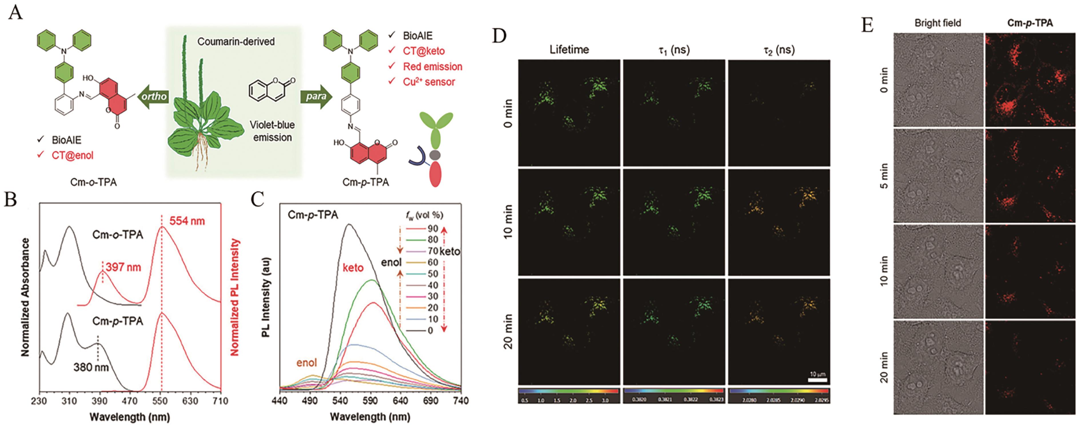
图11 (A) AIE探针Cm-o-TPA和Cm-p-TPA的设计策略; (B)探针Cm-o-TPA和Cm-p-TPA在四氢呋喃(THF)溶液中的归一化吸收光谱; (C)探针Cm-p-TPA在含有不同水分数(fw)的THF/H2O混合溶剂中的荧光光谱; (D)纳米探针Cm-p-TPA NPs(10 μmol/L)在HeLa细胞中的寿命变化; (E)纳米探针Cm-p-TPA NPs(10 μmol/L)对线粒体自噬过程的监测[51]
Fig.11 (A) The design strategy of AIE probe Cm-o-TPA and Cm-p-TPA; (B) The normalized absorption spectra of Cm-o-TPA and Cm-p-TPA in tetrahydrofuran (THF) solution; (C) The fluorescence spectra of Cm-p-TPA in THF/H2O mixed solvent with different water fractions (fw); (D) The lifetime changes of Cm-p-TPA NPs (10 μmol/L) in HeLa cells; (E) Monitoring of mitophagy process with Cm-p-TPA NPs (10 μmol/L)[51]
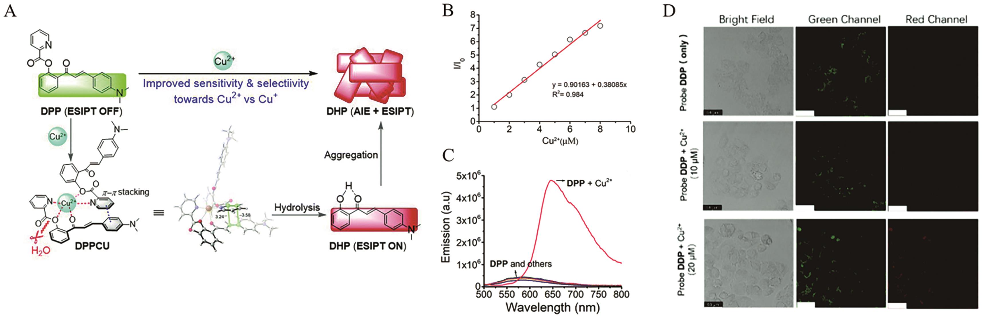
图12 (A) AIE探针DPP的结构及其响应机理; (B)探针DPP的相对荧光强度I648 nm/I0与Cu2+浓度之间的线性关系; (C)探针DPP(20 μmol/L)与不同分析物孵育后的荧光响应; (D)探针DPP(10 μmol/L)在HeLa细胞中的荧光成像[52]
Fig.12 (A) The structure and response mechanism of AIE probe DPP; (B) The linear relationship between the relative fluorescence intensity I648 nm/I0 of probe DPP and the concentration of Cu2+; (C) The fluorescence response of probe DPP (20 μmol/L) after incubation with different analytes; (D) The fluorescence imaging of HeLa cells incubated with probe DPP(10 μmol/L)[52]
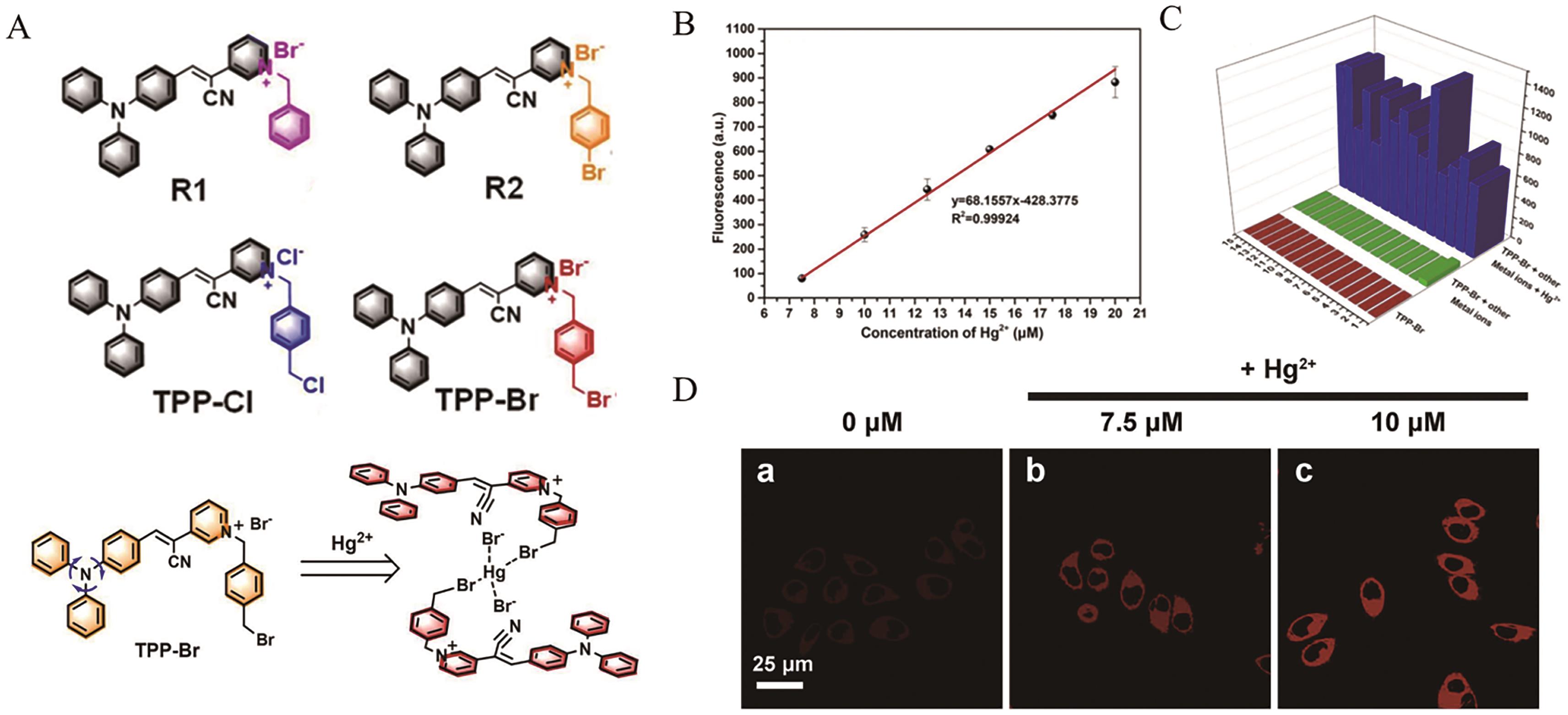
图13 (A) AIE探针R1、R2、TPP-Cl和TPP-Br的结构及TPP-Br的响应机理; (B)探针TPP-Br的荧光强度与Hg2+浓度之间的线性关系; (C)在Hg2+(30?μmol/L)存在的条件下,探针TPP-Br(50 μmol/L)与不同分析物孵育后的荧光响应; (D)探针TPP-Br(10 μmol/L)在HeLa细胞中的荧光成像[57]
Fig.13 (A) The structure of AIE probe R1, R2, TPP-Cl and TPP-Br and response mechanism of TPP-Br; (B) The linear relationship between the fluorescence intensity of probe TPP-Br and the concentration of Hg2+; (C) The fluorescence response of probe TPP-Br (50 μmol/L) after incubation with different analytes in the presence of Hg2+ (30?μmol/L); (D) The fluorescence imaging of HeLa cells incubated with probe TPP-Br (10 μmol/L)[57]
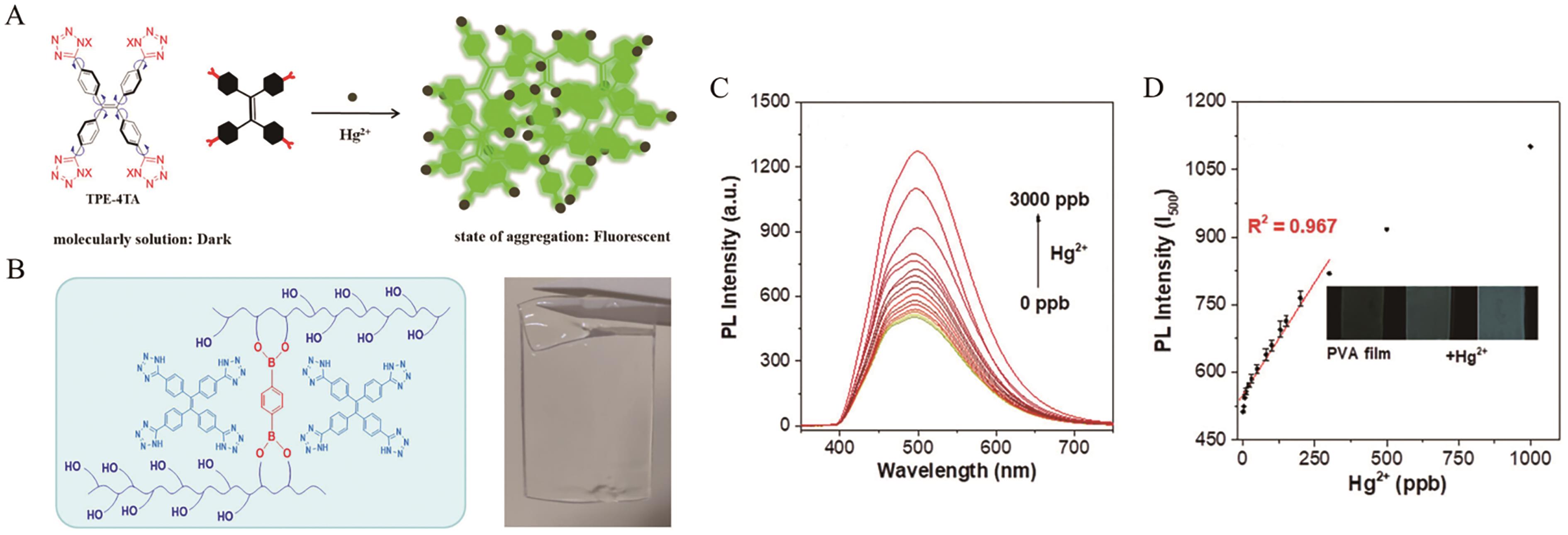
图14 (A) AIE探针TPE-4TA的结构及其响应机理; (B)掺杂TPE-4TA的聚乙烯醇基水凝胶薄膜; (C)聚乙烯醇基水凝胶薄膜与不同浓度Hg2+响应后的荧光光谱; (D)聚乙烯醇基水凝胶薄膜的荧光强度与Hg2+浓度之间的线性关系[58]
Fig.14 (A) The structure and response mechanism of AIE probe TPE-4TA; (B) The polyvinyl alcohol (PVA)-based hydrogel film dopped with TPE-4TA; (C) The fluorescence spectra of PVA-based hydrogel film after reaction with various concentrations of Hg2+; (D) The linear relationship between the fluorescence intensity of PVA-based hydrogel film and the concentration of Hg2+[58]
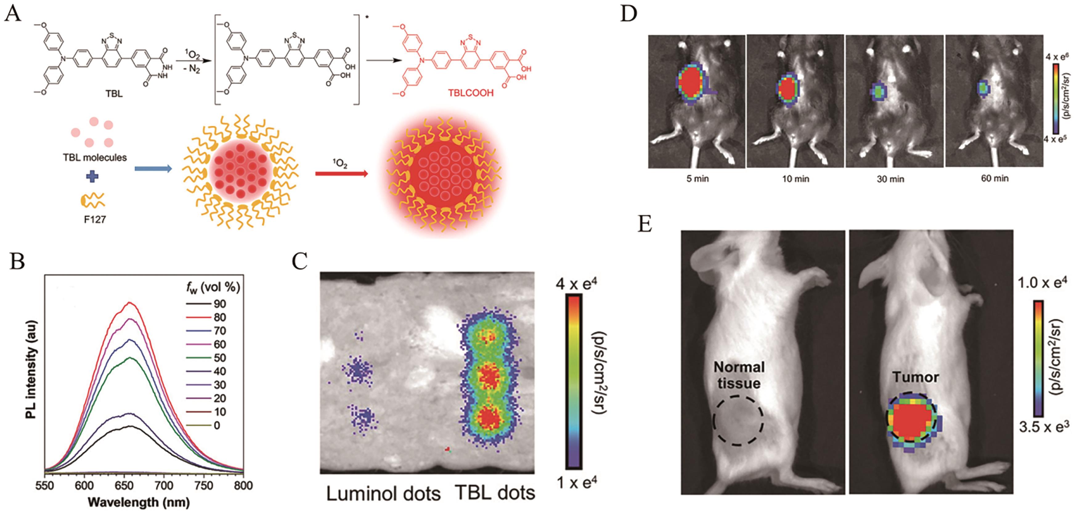
图15 (A) AIE探针TBL的结构及其响应机理; (B)探针TBL在含有不同水分数(fw)的二甲基亚砜(DMSO)/H2O混合溶剂中的荧光光谱; (C)在H2O2/NaClO混合溶液中,由一片约3 mm厚的猪肉火腿覆盖的鲁米诺点(1.2 mmol/L)和TBL点(1.2 mmol/L)的化学发光图像; (D)往小鼠中注射H2O2/NaClO混合溶液后,TBL点的荧光成像; (E) TBL点对小鼠中乳腺肿瘤的化学发光成像[65]
Fig.15 (A) The structure and response mechanism of AIE probe TBL; (B) The fluorescence spectra of TBL in dimethyl sulfoxide (DMSO)/H2O mixed solvent with different water fractions (fw); (C) Chemiluminescence of luminol dots (1.2 mmol/L) and TBL dots (1.2 mmol/L) covered by a piece of approximately 3 mm thick pork ham in a H2O2/NaClO mixed solution; (D) The chemiluminescence imaging of mice after subcutaneous injection of TBL dots with H2O2 and NaClO; (E) The chemiluminescence imaging of breast tumor-bearing mice after injection of TBL dots[65]
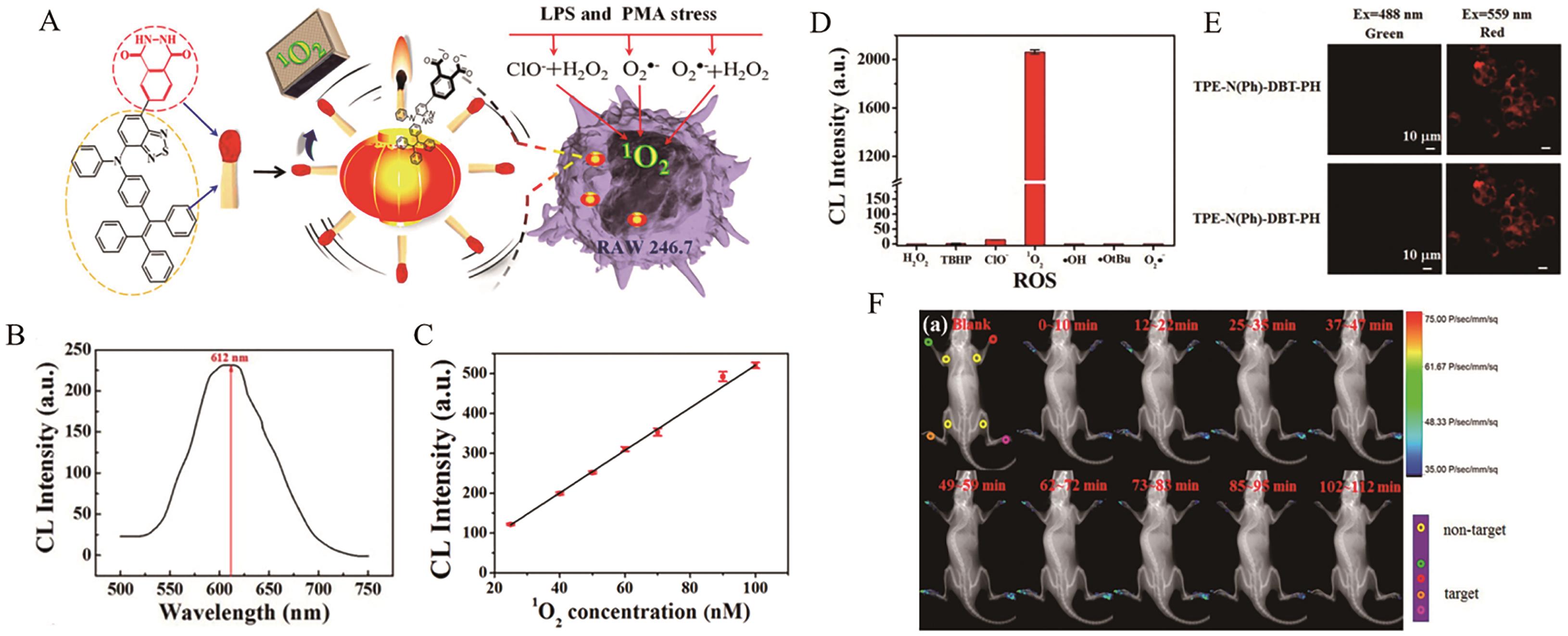
图16 (A) AIE探针TPE-N(Ph)-DBT-PH的结构及其响应机理; (B)探针TPE-N(Ph)-DBT-PH的化学发光光谱; (C)探针TPE-N(Ph)-DBT-PH的化学发光强度与1O2浓度之间的线性关系; (D)探针TPE-N(Ph)-DBT-PH(10 μmol/L)与不同分析物孵育后的化学发光响应; (E)探针TPE-N(Ph)-DBT-PH在RAW 264.7细胞中的化学发光成像; (F)探针TPE-N(Ph)-DBT-PH在关节炎小鼠模型中的化学发光成像[66]
Fig.16 (A) The structure and response mechanism of AIE probe TPE-N(Ph)-DBT-PH; (B) The chemiluminescence spectra of TPE-N(Ph)-DBT-PH; (C) The linear relationship between the chemiluminescence intensity of probe TPE-N(Ph)-DBT-PH and the concentration of 1O2; (D) The chemiluminescence response of probe TPE-N(Ph)-DBT-PH (10 μmol/L) after incubation with different analytes; (E) The chemiluminescence imaging of RAW 264.7 cells incubated with probe TPE-N(Ph)-DBT-PH; (F) The chemiluminescence imaging of a arthritismouse model after injection of TPE-N(Ph)-DBT-PH[66]
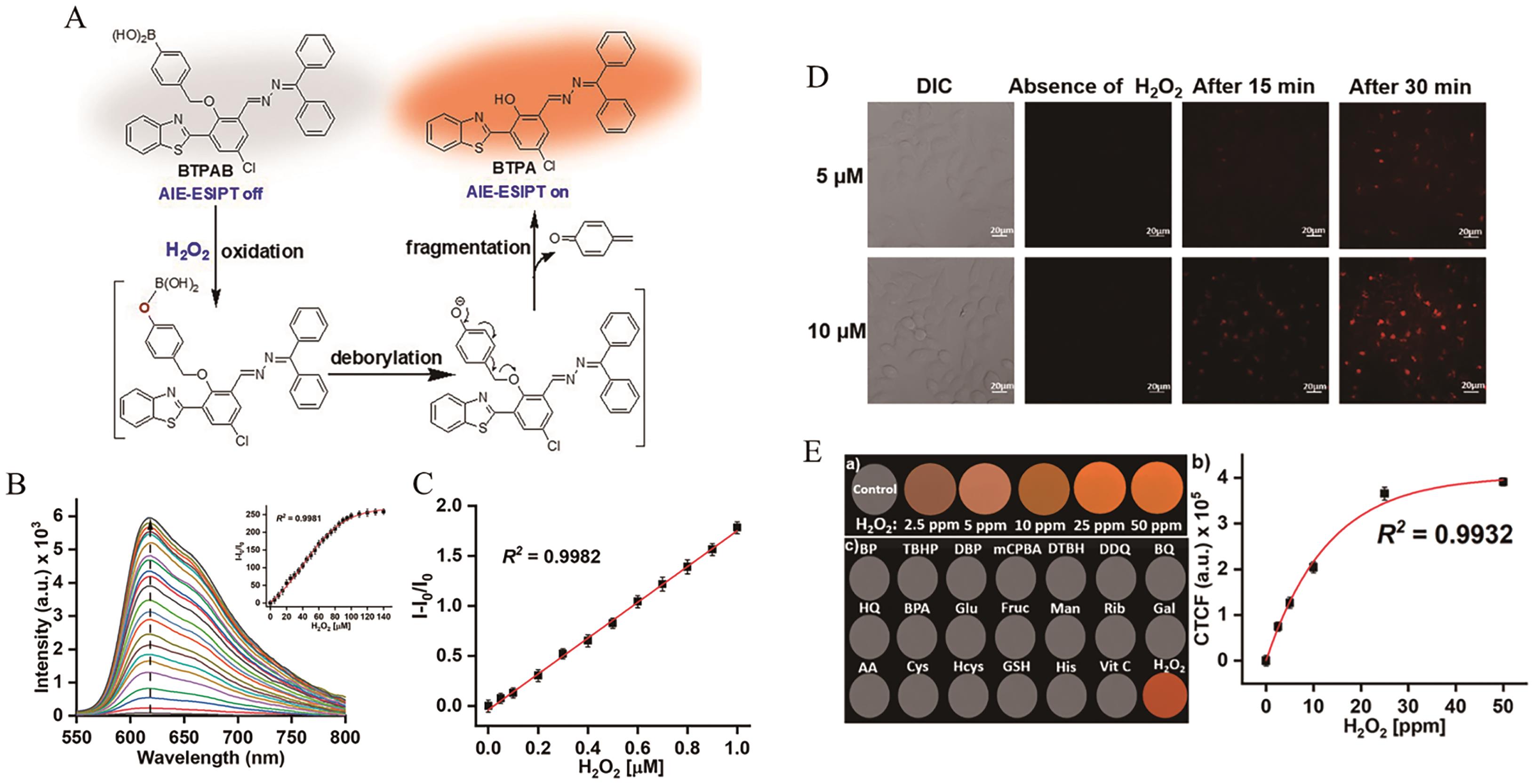
图17 (A) AIE探针BTPAB的结构及其响应机理; (B)探针BTPAB(10 μmol/L)与不同浓度H2O2响应后的荧光光谱; (C)探针BTPAB的相对荧光强度与H2O2浓度之间的线性关系; (D)探针BTPAB在HeLa细胞中的荧光成像; (E)探针BTPAB在TLC板上的荧光响应图像及相应的线性关系[69]
Fig.17 (A) The structure and response mechanism of AIE probe BTPAB; (B) The fluorescence spectra of BTPAB(10 μmol/L) after reaction with various concentrations of H2O2; (C) The linear relationship between the fluorescence intensity of BTPAB and the concentration of H2O2; (D) The fluorescence imaging of HeLa cells incubated with probe BTPAB; (E) Fluorescence response of BTPAB to H2O2 on TLC plates and corresponding linear relationship[69]
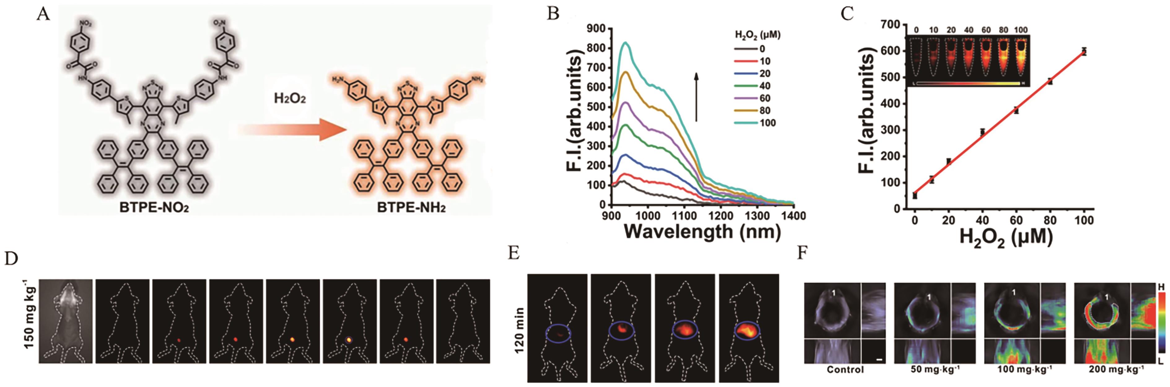
图18 (A) AIE探针BTPE-NO2的结构及其响应机理; (B)探针BTPE-NO2@F127(BTPE-NO2 32.6?μg/mL)与不同浓度H2O2响应后的荧光光谱; (C)探针BTPE-NO2@F127的荧光强度与H2O2浓度之间的线性关系; (D) BTPE-NO2@F127在间质性膀胱炎小鼠模型中的荧光成像; (E) BTPE-NO2@F127在曲唑酮诱导的肝损伤小鼠模型中的荧光成像; (F) BTPE-NO2@F127在曲唑酮诱导的肝损伤小鼠模型中的多光谱光声断层扫描成像[70]
Fig.18 (A) The structure and response mechanism of AIE probe BTPE-NO2; (B) The fluorescence spectra of BTPE-NO2@F127 (BTPE-NO2 32.6?μg/mL) after reaction with various concentrations of H2O2; (C) The linear relationship between the fluorescence intensity of BTPE-NO2@F127 and the concentration of H2O2; (D) The fluorescence imaging of an interstitial cystitis mouse model after injection of BTPE-NO2@F127; (E) The fluorescence imaging of a trazodone-induced liver injury mouse model after injection of BTPE-NO2@F127; (F) The multispectral optoacoustic tomography imaging of a trazodone-induced liver injury mouse model after injection of BTPE-NO2@F127[70]
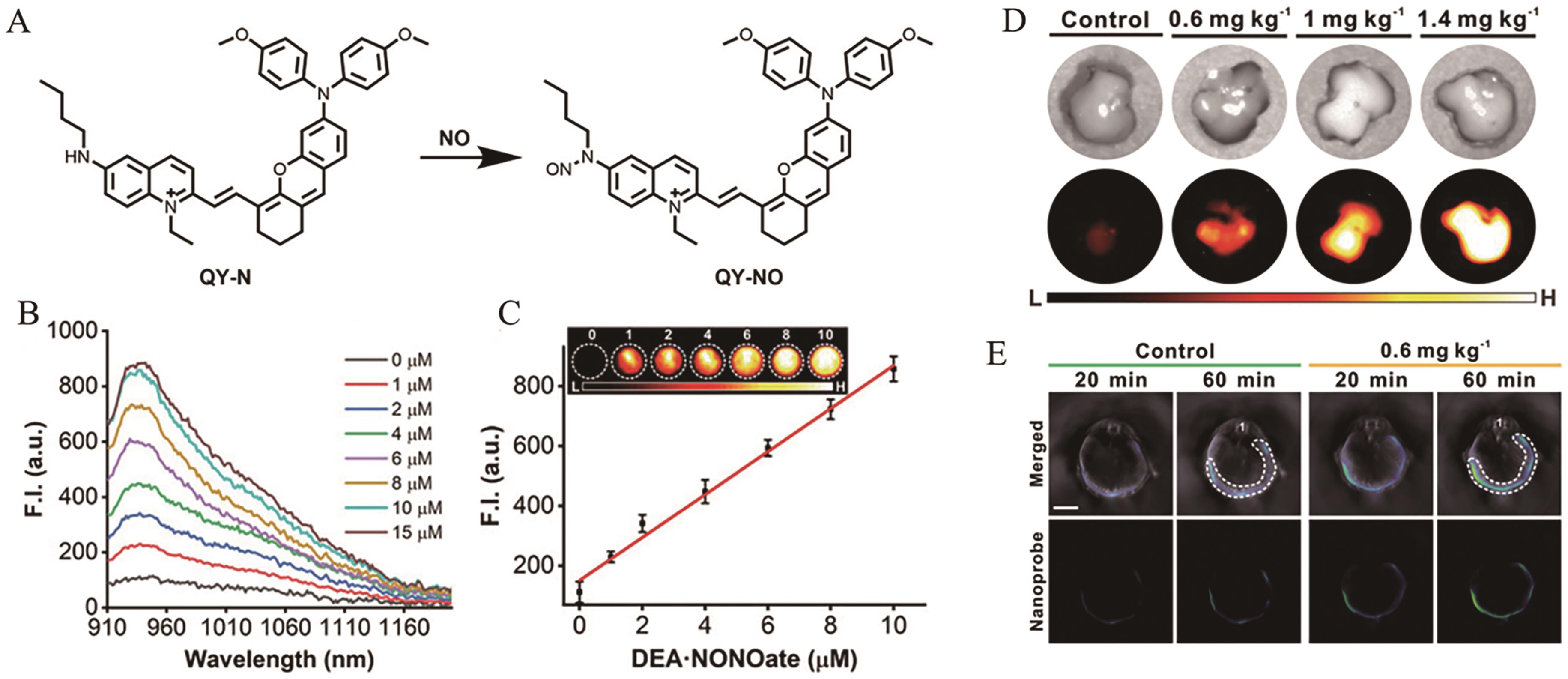
图19 (A) AIE探针QY-N的结构及其响应机理;(B)探针QY-N(10 μmol/L)与不同浓度NO响应后的荧光光谱; (C)探针QY-N的荧光强度与NO浓度之间的线性关系; (D) QY-N在小鼠肝脏中的荧光成像; (E) QY-N在小鼠肝脏中的多光谱光声断层扫描成像[73]
Fig.19 (A) The structure and response mechanism of AIE probe QY-N; (B) The fluorescence spectra of QY-N (10 μmol/L) after reaction with various concentrations of NO; (C) The linear relationship between the fluorescence intensity of QY-N and the concentration of NO; (D) The fluorescence imaging of livers incubated with probe QY-N; (E) The multispectral optoacoustic tomography imaging of livers after injection of QY-N[73]
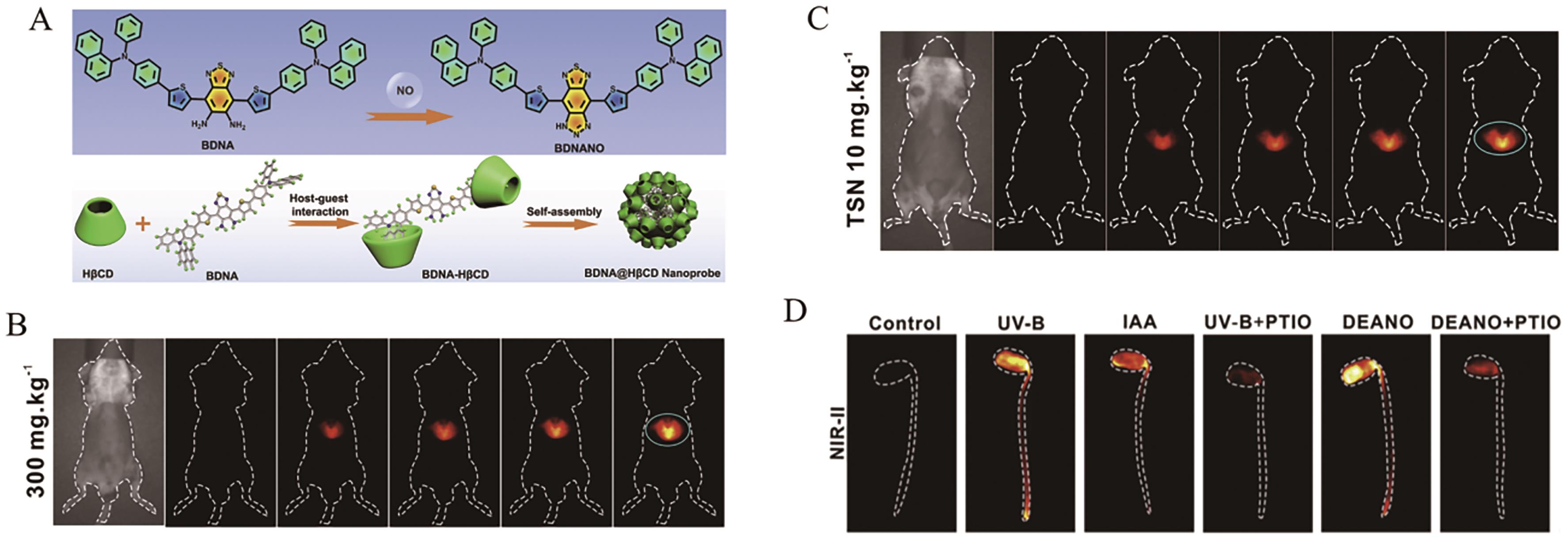
图20 (A)探针BNDA的结构及其响应机理; (B) BNDA@HβCD在乙酰氨基酚诱导的肝损伤小鼠模型中的荧光成像; (C) BNDA@HβCD在川楝素诱导的肝损伤小鼠模型中的荧光成像; (D) BNDA@HβCD在豆芽中的荧光成像[74]
Fig.20 (A) The structure and response mechanism of AIE probe BNDA; (B) The fluorescence imaging of an APAP-induced liver injury mouse model after injection of BNDA@HβCD; (C) The fluorescence imaging of an TSN-induce dliver injury mouse model after injection of BNDA@HβCD; (D) The fluorescence imaging of soybean sprouts[74]

图21 (A) AIE探针DNBS-HCA的结构及其响应机理; (B)探针DNBS-HCA分别与GSH、Cys和Hcy响应后的荧光光谱; (C)探针DNBS-HCA的荧光强度与GSH、Cys和Hcy浓度之间的线性关系; (D)探针DNBS-HCA在PC-3细胞和A549细胞中的荧光成像; (E)探针DNBS-HCA在试纸上对Cys的荧光响应图像[77]
Fig.21 (A) The structure and response mechanism of AIE probe DNBS-HCA; (B) The fluorescence spectra of DNBS-HCA after reaction with various concentrations of GSH, Cys and Hcy, repectively; (C) The linear relationship between the fluorescence intensity of DNBS-HCA and the concentration of GSH, Cys and Hcy, repectively; (D) The fluorescence imaging of PC-3 cells and A549 cells incubated with probe DNBS-HCA; (E) Fluorescence response of DNBS-HCA to Cys on test papers[77]
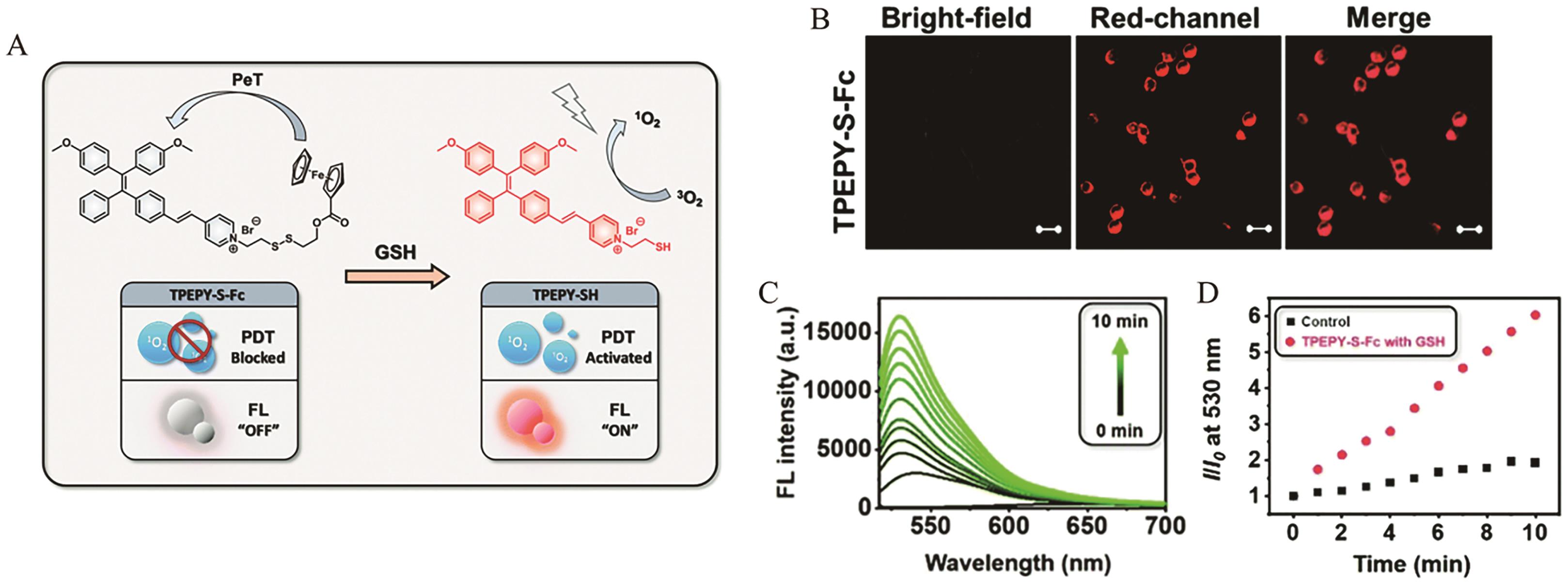
图22 (A) AIE探针TPEPY-S-Fc的结构及其响应机理; (B)探针TPEPY-S-Fc在CT-26细胞中的荧光成像; (C)在TPEPY-S-Fc(20 μmol/L)和GSH(200 μmol/L)存在的条件下,单线态氧探针SOSG的荧光光谱; (D) SOSG的相对荧光强度I530 nm/I0与时间的关系[78]
Fig.22 (A) The structure and response mechanism of AIE probe TPEPY-S-Fc; (B) The fluorescence imaging of CT-26 cells incubated with probe TPEPY-S-Fc; (C) The fluorescence spectra of 1O2 probe SOSG in the presence of TPEPY-S-Fc (20 μmol/L) and GSH (200 μmol/L); (D) The time-dependent relative fluorescence intensity I530 nm/I0 of probe SOSG[78]
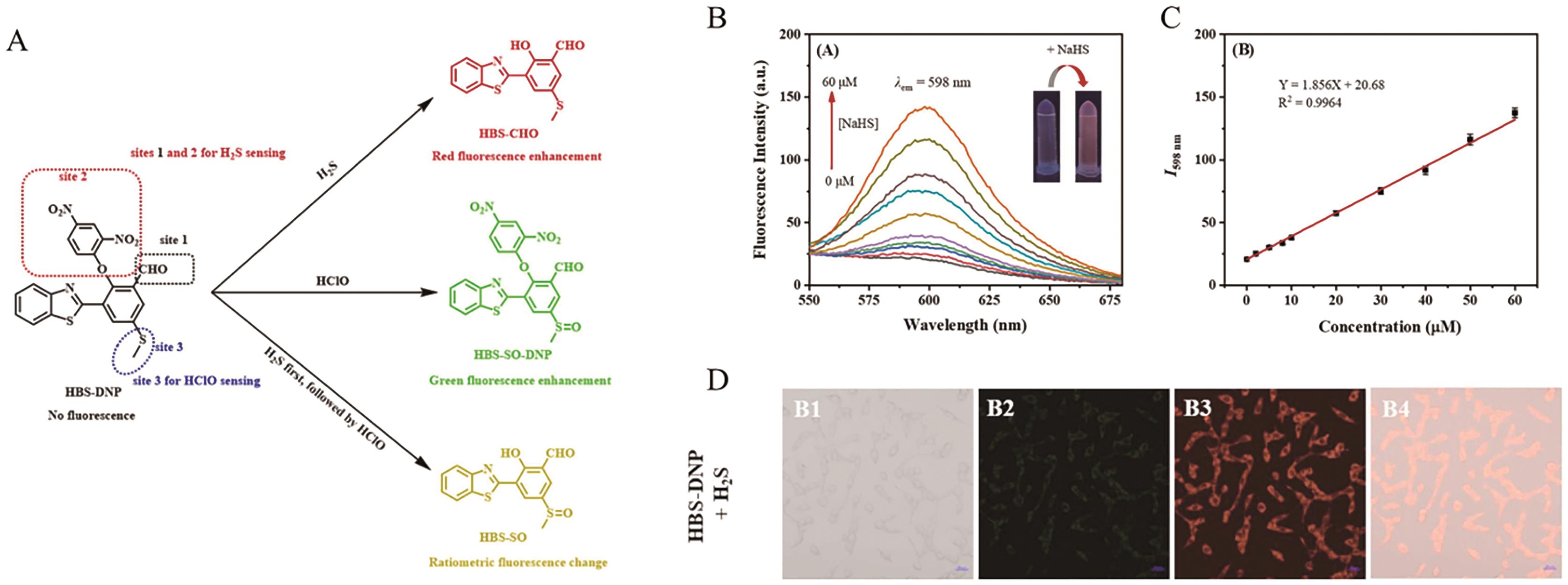
图23 (A) AIE探针HBS-DNP的结构及其响应机理; (B)探针HBS-DNP(10 μmol/L)与不同浓度H2S响应后的荧光光谱; (C)探针HBS-DNP的荧光强度与H2S浓度之间的线性关系; (D) HBS-DNP(10 μmol/L)在Hela细胞中的荧光成像[82]
Fig.23 (A) The structure and response mechanism of AIE probe HBS-DNP; (B) The fluorescence spectra of HBS-DNP (10 μmol/L) after reaction with various concentrations of H2S; (C) The linear relationship between the fluorescence intensity of HBS-DNP and the concentration of H2S; (D) The fluorescence imaging of Hela cells incubated with probe HBS-DNP(10 μmol/L)[82]
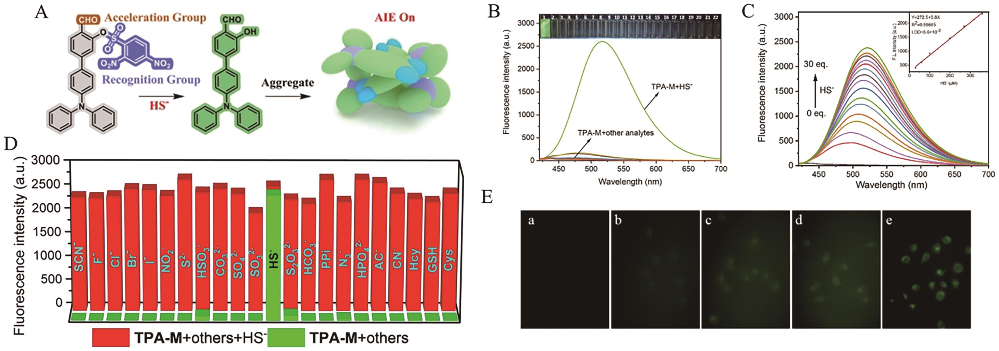
图24 (A) AIE探针TPA-M的结构及其响应机理; (B) TPA-M(10 μmol/L)与不同分析物孵育后的荧光响应; (C)探针TPA-M(10 μmol/L)与不同浓度H2S响应后的荧光光谱及相应的线性关系; (D)在H2S(300?μmol/L)存在的条件下,探针TPA-M(10 μmol/L)与不同分析物孵育后的荧光响应; (E)探针TPA-M在MCF-7细胞中的荧光成像[83]
Fig.24 (A) The structure and response mechanism of AIE probe TPA-M; (B) The fluorescence response of probe TPA-M (10 μmol/L) after incubation with different analytes; (C) The fluorescence spectra of TPA-M (10 μmol/L) after reaction with various concentrations of H2S and corresponding linear relationship; (D) The fluorescence response of probe TPA-M (10 μmol/L) after incubation with different analytes in the presence of H2S(300?μmol/L); (E) The fluorescence imaging of MCF-7 cells incubated with probe TPA-M[83]
| [1] | GRIFFITHS H R, MØLLER L, BARTOSZ G, et al. Biomarkers[J]. Mol Asp Med, 2022, 23: 101-208. |
| [2] | GU X, KWOK R T K, LAM J W Y, et al. AIEgens for biological process monitoring and disease theranostics[J]. Biomaterials, 2017, 146: 115-135. |
| [3] | CULLEN N C, LEUZY A, JANELIDZE S, et al. Plasma biomarkers of Alzheimer′s disease improve prediction of cognitive decline in cognitively unimpaired elderly populations[J]. Nat Commun, 2021, 12(1): 3555-3563. |
| [4] | BROZA Y Y, ZHOU X, YUAN M, et al. Disease detection with molecular biomarkers: from chemistry of body fluids to nature-inspired chemical sensors[J]. Chem Rev, 2019, 119(22): 11761-11817. |
| [5] | KIM K, LEE C H, PARK C B. Chemical sensing platforms for detecting trace-level Alzheimer's core biomarkers[J]. Chem Soc Rev, 2020, 49(15): 5446-5472. |
| [6] | WHITFIELD M L, GEORGE L K, GRANT G D, et al. Common markers of proliferation[J]. Nat Rev Cancer, 2006, 6(2): 99-106. |
| [7] | ZHOU Y, TAO L, QIU J, et al. Tumor biomarkers for diagnosis, prognosis and targeted therapy[J]. Sig Transduct Target Ther, 2024, 9(1): 132-217. |
| [8] | WEISSLEDER R, PITTET M J. Imaging in the era of molecular oncology[J]. Nature, 2008, 452(7187): 580-589. |
| [9] | WEISSLEDER R, NAHRENDORF M. Advancing biomedical imaging[J]. Proc Natl Acad Sci, 2015, 112(47): 14424-14428. |
| [10] | HERREMANS E, MELADO-HERREROS A, DEFRAEYE T, et al. Comparison of X-ray CT and MRI of watercore disorder of different apple cultivars[J]. Postharvest Biol Tec, 2014, 87: 42-50. |
| [11] | QU H, FAN C, CHEN M, et al. Recent advances of fluorescent biosensors based on cyclic signal amplification technology in biomedical detection[J]. J Nanobiotechnol, 2021, 19(1): 403-430. |
| [12] | TANG Y, PEI F, LU X, et al. Recent advances on activatable NIR‐Ⅱ fluorescence probes for biomedical imaging[J]. Adv Opt Mater, 2019, 7(21): 1900917. |
| [13] | WANG S, LI X, CHONG S Y, et al. In vivo three‐photon imaging of lipids using ultrabright fluorogens with aggregation‐induced emission[J]. Adv Mater, 2021, 33(11): 20200749. |
| [14] | XIAO D, QI H, TENG Y, et al. Advances and challenges of fluorescent nanomaterials for synthesis and biomedical applications[J]. Nanoscale Res Lett, 2021, 16(1): 167-179. |
| [15] | XU Y, XU R, WANG Z, et al. Recent advances in luminescent materials for super-resolution imaging via stimulated emission depletion nanoscopy[J]. Chem Soc Rev, 2021, 50(1): 667-690. |
| [16] | GOPIKRISHNA P, MEHER N, IYER P K. Functional 1,8-naphthalimide AIE/AIEEgens: recent advances and prospects[J]. ACS Appl Mater Interfaces, 2017, 10(15): 12081-12111. |
| [17] | XU L, JIANG X, LIANG K, et al. Frontier luminous strategy of functional silica nanohybrids in sensing and bioimaging: from ACQ to AIE[J]. Aggregate, 2021, 3(1): e121-e149. |
| [18] | LI D, YU J. AIEgens‐functionalized inorganic‐organic hybrid materials: fabrications and applications[J]. Small, 2016, 12(47): 6478-6494. |
| [19] | BEDDARD G S, PORTER G. Concentration quenching in chlorophyll[J]. Nature, 1976, 260: 366-367. |
| [20] | LUO J, XIE Z, LAM J W Y, et al. Aggregation-induced emission of 1-methyl-1,2,3,4,5-pentaphenylsilole[J]. Chem Commun, 2001, 18: 1740-1741. |
| [21] | TU Y, ZHAO Z, LAM J W Y, et al. Mechanistic connotations of restriction of intramolecular motions (RIM)[J]. Natl Sci Rev, 2021, 8(6): nwaa260. |
| [22] | ZHANG J, ZHANG H, LAM J W Y, et al. Restriction of intramolecular motion(RIM): investigating AIE mechanism from experimental and theoretical studies[J]. Chem Res Chin Univ, 2021, 37(1): 1-15. |
| [23] | WU Q, LI Y, WANG L, et al. Aggregation-induced emission: an emerging concept in brain science[J]. Biomaterials, 2022, 286: 121581. |
| [24] | ZHAO Y Q, YU L, ZHANG L, et al. Activated aggregation‐induced emission therapeutics agents for triggering regulated cell death[J]. Aggregate, 2024, 5(3): e503-e530. |
| [25] | WANG H, LI Q, ALAM P, et al. Aggregation-induced emission (AIE), life and health[J]. ACS Nano, 2023, 17(15): 14347-14405. |
| [26] | REN K, ZHANG B, GUO J, et al. Aggregation-induced emission (AIE) for next-generation biosensing and imaging: a review[J]. Biosens Bioelectron, 2025, 271: 117067. |
| [27] | GONG S, QIN A, ZHANG Y, et al. A new ratiometric AIE fluorescent probe for detecting cysteine in food samples and imaging in the biological system[J]. Food Chem, 2023, 400: 134108. |
| [28] | ZHAO H, LI N, MA C, et al. An AIE probe for long-term plasma membrane imaging and membrane-targeted photodynamic therapy[J]. Chin Chem Lett, 2023, 34(4): 107699. |
| [29] | DUO Y, LUO G, ZHANG W, et al. Noncancerous disease-targeting AIEgens[J]. Chem Soc Rev, 2023, 52(3): 1024-1067. |
| [30] | ZHANG J, CHAI X, HE X P, et al. Fluorogenic probes for disease-relevant enzymes[J]. Chem Soc Rev, 2019, 48(2): 683-722. |
| [31] | SHARMA U, PAL D, PRASAD R. Alkaline phosphatase: an overview[J]. Ind J Clin Biochem, 2013, 29(3): 269-278. |
| [32] | SMITH B A H, BERTOZZI C R. The clinical impact of glycobiology: targeting selectins, siglecs and mammalian glycans[J]. Nat Rev Drug Discovery, 2021, 20(3): 217-243. |
| [33] | RADER B A. Alkaline phosphatase, an unconventional immune protein[J]. Front Immunol, 2017, 8: 897-902. |
| [34] | LAM K W K, CHAU J H C, YU E Y, et al. An alkaline phosphatase-responsive aggregation-induced emission photosensitizer for selective imaging and photodynamic therapy of cancer cells[J]. ACS Nano, 2023, 17(8): 7145-7156. |
| [35] | LI H, YAO Q, XU F, et al. An activatable AIEgen probe for high-fidelity monitoring of overexpressed tumor enzyme activity and its application to surgical tumor excision[J]. Angew Chem Int Ed, 2020, 59(25): 10186-10195. |
| [36] | CAI Y, ZHOU H, ZHU Y, et al. Elimination of senescent cells by β-galactosidase-targeted prodrug attenuates inflammation and restores physical function in aged mice[J]. Cell Res, 2020, 30(7): 574-589. |
| [37] | GU K, QIU W, GUO Z, et al. An enzyme-activatable probe liberating AIEgens: on-site sensing and long-term tracking of β-galactosidase in ovarian cancer cells[J]. Chem Sci, 2019, 10(2): 398-405. |
| [38] | FAN F, ZHANG L, ZHOU X, et al. A sensitive fluorescent probe for β-galactosidase activity detection and application in ovarian tumor imaging[J]. J Mater Chem B, 2021, 9(1): 170-175. |
| [39] | TIAN H, LIN W, HU XL, et al. Ratiometric sensing of β-galactosidase based on excited-state intramolecular proton transfer (ESIPT) and solid-state luminescence enhancement[J]. Org Chem Front, 2023, 10(12): 2913-2917. |
| [40] | FENG B, CHU F, HUANG X, et al. Debut of a NIR ESIPT-based fluorescent probe with synergistic effects for boosting high-contrast imaging of β-galactosidase in ovarian cancer[J]. Sens Actuators B, 2023, 396: 134541. |
| [41] | OUYANG J, SUN L, ZENG Z, et al. Nanoaggregate probe for breast cancer metastasis through multispectral optoacoustic tomography and aggregation‐induced NIR-Ⅰ/Ⅱ fluorescence imaging[J]. Angew Chem Int Ed, 2019, 59(25): 10111-10121. |
| [42] | SHEN D, DING S, LU Q, et al. Nitroreductase-responsive fluorescent “off-on” photosensitizer for hypoxic tumor imaging and dual-modal therapy[J]. ACS Omega, 2024, 9(28): 30685-30697. |
| [43] | VALLEE B L, FALCHUK K H. The biochemical basis of zinc physiology[J]. Physiol Rev, 1993, 73: 79-118. |
| [44] | CHEN X, CAI Q, LIANG R, et al. Copper homeostasis and copper-induced cell death in the pathogenesis of cardiovascular disease and therapeutic strategies[J]. Cell Death Dis, 2023, 14(2): 105-116. |
| [45] | HAMA A K H, MUSTAFA F S, OMER K M, et al. Heavy metal pollution in the aquatic environment: efficient and low-cost removal approaches to eliminate their toxicity: a review[J]. RSC Adv, 2023, 13(26): 17595-17610. |
| [46] | FREDERICKSON C J, KOH J Y, BUSH A I. The neurobiology of zinc in health and disease[J]. Nat Rev Neurosci, 2005, 6(6): 449-462. |
| [47] | XU J, XIONG J, QIN Y, et al. A novel quinolinyl-tetraphenylethene-based fluorescence “turn-on” sensor for Zn2+ with a large stokes shift and its applications for portable test strips and biological imaging[J]. Mater Chem Front, 2020, 4(11): 3338-3348. |
| [48] | JOY F, CHAITHRA K P, NIZAM A, et al. A Multi-Stimuli responsive organic luminogen with aggregation induced emission for the selective detection of Zn2+ ions in solution and solid state[J]. Chem Eng J, 2023, 453: 139798. |
| [49] | GARZA N M, SWAMINATHAN A B, MAREMANDA K P, et al. Mitochondrial copper in human genetic disorders[J]. Trends Endocrinol Metab, 2023, 34(1): 21-33. |
| [50] | RAMCHANDANI D, BERISA M, TAVAREZ D A, et al. Copper depletion modulates mitochondrial oxidative phosphorylation to impair triple negative breast cancer metastasis[J]. Nat Commun, 2021, 12(1): 7311-7326. |
| [51] | CAI X M, LI S, WANG W J, et al. Natural acceptor of coumarin-isomerized red-emissive bioAIEgen for monitoring Cu2+ concentration in live cells via FLIM[J]. Adv Sci, 2023, 11(9): 2307078. |
| [52] | JIANG J, SUN H, HU Y, et al. AIE+ESIPT activity-based NIR Cu2+ sensor with dye participated binding strategy[J]. Chem Commun, 2021, 57(62): 7685-7688. |
| [53] | NIU X, ZHANG H, WU X, et al. An AIE-active “turn-off” fluorescent sensor for highly selective and sensitive detection of Cu2+ ions[J]. J Mol Struct, 2022, 1264: 133294. |
| [54] | NESCI S, TROMBETTI F, PIRINI M, et al. Mercury and protein thiols: stimulation of mitochondrial F1FO-ATPase and inhibition of respiration[J]. Chem Biol Interact, 2016, 260: 42-49. |
| [55] | KIRAN, BHARTI R, SHARMA R. Effect of heavy metals: an overview[J]. Mater Today: Proc, 2022, 51: 880-885. |
| [56] | TONAZZI A, GIANGREGORIO N, CONSOLE L, et al. Mitochondrial carnitine/acylcarnitine transporter, a novel target of mercury toxicity[J]. Chem Res Toxicol, 2015, 28(5): 1015-1022. |
| [57] | TAN T, ZHANG C, HAN Y, et al. Fine-tuning bromide AIE probes for Hg2+ detection in mitochondria with wash-free staining[J]. J Hazard Mater, 2024, 464: 132999. |
| [58] | WU S, YANG Y, CHENG Y, et al. Fluorogenic detection of mercury ion in aqueous environment using hydrogel-based AIE sensing films[J]. Aggregate, 2022, 4(3): e287-e294. |
| [59] | WALLACE D C. Mitochondrial diseases in man and mouse[J]. Science, 1999, 283: 1482-1488. |
| [60] | YANG B, CHEN Y, SHI J. Reactive oxygen species (ROS)-based nanomedicine[J]. Chem Rev, 2019, 119(8): 4881-4985. |
| [61] | GRIFFITHS H R, GAO D, PARARASA C. Redox regulation in metabolic programming and inflammation[J]. Redox Biol, 2017, 12: 50-57. |
| [62] | BEGHETTO C, RENKEN C, ERIKSSON O, et al. Implications of the generation of reactive oxygen species by photoactivated calcein for mitochondrial studies[J]. Eur J Biochem, 2000, 267: 5585-5592 |
| [63] | RYTER S W, TYRRELL R M. Singlet molecular oxygen (1O2) a possible effector of eukaryotic gene expression[J]. Free Radic Biol Med, 1998, 24: 1520-1534. |
| [64] | KLOTZ L O, BRIVIBA K, SIES H. Mitogen-activated protein kinase activation by singlet oxygen and ultraviolet A[J]. Methods Enzymol, 2000, 319: 130-143. |
| [65] | LIU C, WANG X, LIU J, et al. Near‐infrared AIE dots with chemiluminescence for deep-tissue imaging[J]. Adv Mater, 2020, 32(43): 2004685. |
| [66] | LYU J, CHENG M, LIU J, et al. An aggregation-induced emission nanosensor for real-time chemiluminescent sensing of light-independent intracellular singlet oxygen[J]. ACS Appl Mater Interfaces, 2022, 14(48): 54081-54089. |
| [67] | RHEE S G. H2O2, a necessary evil for cell signaling[J]. Science, 2006, 312: 1882-1883. |
| [68] | FINKEL T, HOLBROOK N J. Oxidants, oxidative stress and the biology of ageing[J]. Nature, 2000, 408: 239-247. |
| [69] | BHOSLE A A, BANERJEE M, GUPTA V, et al. Mechanochemical synthesis of an AIE-TICT-ESIPT active orange-emissive chemodosimeter for selective detection of hydrogen peroxide in aqueous media and living cells, and solid-phase quantitation using a smartphone[J]. New J Chem, 2022, 46(39): 18961-18972. |
| [70] | CHEN J, CHEN L, WU Y, et al. A H2O2-activatable nanoprobe for diagnosing interstitial cystitis and liver ischemia-reperfusion injury via multispectral optoacoustic tomography and NIR-Ⅱ fluorescent imaging[J]. Nat Commun, 2021, 12(1): 6870-6884. |
| [71] | IWAKIRI Y, KIM M Y. Nitric oxide in liver diseases[J]. Trends Pharmacol Sci, 2015, 36(8): 524-536. |
| [72] | JIN G, GAO Z, LIU Y, et al. Polymeric nitric oxide delivery nanoplatforms for treating cancer, cardiovascular diseases, and infection[J]. Adv Healthcare Mater, 2020, 10(3): 2001550. |
| [73] | SUN L, OUYANG J, MA Y, et al. An activatable probe with aggregation‐induced emission for detecting and imaging herbal medicine induced liver injury with optoacoustic imaging and NIR‐II fluorescence imaging[J]. Adv Healthcare Mater, 2021, 10(24): 2100867. |
| [74] | CHEN J, CHEN L, FANG Y, et al. Refashioning benzothiadiazole dye as an activatable nanoprobe for biomarker detection with NIR-Ⅱ fluorescence/optoacoustic imaging[J]. Cell Reports Phys Sci, 2022, 3(2): 100570. |
| [75] | FILOMENI G, ROTILIO G, CIRIOLO M R. Cell signalling and the glutathione redox system[J]. Biochem Pharmacol, 2002, 64(5/6): 1057-1064. |
| [76] | TOWNSEND D M, TEW K D, TAPIERO H. The importance of glutathione in human disease[J]. Biomed Pharmacother, 2003, 57(3/4): 145-155. |
| [77] | DAI F, ZHAO M, YANG F, et al. An ESIPT coupled AIE fluorescent probe for biothiols detection and imaging based on a chalcone fluorophore[J]. Dyes Pigm, 2020, 183: 108627. |
| [78] | ZHANG Y H, LI X, HUANG L, et al. AIE based GSH activatable photosensitizer for imaging-guided photodynamic therapy[J]. Chem Commun, 2020, 56(71): 10317-10320. |
| [79] | SHEN Y, WEI Y, GAO X, et al. Engineering an enzymatic cascade catalytic smartphone-based sensor for onsite visual ratiometric fluorescence-colorimetric dual-mode detection of methyl mercaptan[J]. Environ Sci Technol, 2023, 57(4): 1680-1691. |
| [80] | LEE M, SCHWAB C, YU S, et al. Astrocytes produce the antiinflammatory and neuroprotective agent hydrogen sulfide[J]. Neurobiol Aging, 2009, 30(10): 1523-1534. |
| [81] | YANG G, WU L, JIANG B, et al. H2S as a physiologic vasorelaxant hypertension in mice with deletion of cystathionine γ-lyase[J]. Science, 2008, 322: 587-590. |
| [82] | GU B, DAI C, ZHOU Z, et al. Rational construction of an AIE-active fluorescent probe bearing three reaction sites for individual and continuous detection of H2S and HClO with single-wavelength excitation[J]. Sens Actuators, B, 2023, 375: 132900-132907. |
| [83] | MENG Y, YAO X, ZHONG K, et al. An aggregation-induced emission‐based fluorescence turn-on probe for efficient detection of HS- in water, wine, and living cells[J]. Eur J Org Chem, 2023, 26(18): e202300022. |
| [1] | 孟真真, 杨金凤, 延钰康. 苯并吡喃腈类近红外聚集诱导荧光探针在粘度检测及细胞成像中的应用[J]. 应用化学, 2025, 42(7): 971-981. |
| [2] | 王振操, 吕宁, 陈梁芳, 饶跃峰. 聚集诱导发光分子用于荧光成像治疗研究进展[J]. 应用化学, 2025, 42(6): 741-756. |
| [3] | 李玲, 唐俐婷, 杨君洁, 闫力强, 李建平. 有机小分子荧光探针对肼检测的研究进展[J]. 应用化学, 2025, 42(4): 453-465. |
| [4] | 王松柏, 李天宇, 魏梦姝, 郭炘明, 李俊芬, 周影, 彭秀英, 董川. 基于硅量子点荧光法和比色法双模式检测绿原酸[J]. 应用化学, 2025, 42(4): 511-521. |
| [5] | 肖巨颖, 赵霞, 林园, 苏朝晖. 功能型聚乙二醇化壳聚糖纳米颗粒的制备和表征[J]. 应用化学, 2025, 42(3): 386-395. |
| [6] | 高长江, 黄鑫, 田亚洋, 余海峰, 王小波. 一种近红外快速响应型硫化氢荧光探针的合成及细胞成像[J]. 应用化学, 2025, 42(2): 192-200. |
| [7] | 许良, 刘健鹏, 青建. 有机八极分子双光子激发荧光材料的设计合成与性能的研究进展[J]. 应用化学, 2025, 42(1): 29-41. |
| [8] | 尹娜, 王樱蕙, 张洪杰. 稀土纳米材料在脑肿瘤成像和治疗中的研究进展[J]. 应用化学, 2024, 41(3): 309-327. |
| [9] | 铁德金, 许美佳, 唐晓丹, 贾宏敏, 于洪梅. 一种可用于检测pH和H2O的双比率荧光探针[J]. 应用化学, 2024, 41(10): 1491-1501. |
| [10] | 赵永梅, 程沛璇, 谢京娇, 罗稳. 基于激发态分子内质子转移机制的3-羟基黄酮探针用于检测生物硫醇[J]. 应用化学, 2024, 41(10): 1502-1510. |
| [11] | 赵欣雨, 秦作佳, 张晓兵, 袁林. 近红外二区激活型小分子荧光探针研究进展[J]. 应用化学, 2024, 41(1): 39-59. |
| [12] | 袁丽珍, 林妮娅, 张云帆, 胡晶晶, 娄筱叮, 夏帆. 基于外表面修饰的固态纳米通道用于生物标志物分析的研究进展[J]. 应用化学, 2024, 41(1): 87-99. |
| [13] | 仝大伟, 孔明, 向育斌. 含甲氧基四苯乙烯咪唑化合物的合成、光物理性质、理论计算及细胞成像[J]. 应用化学, 2023, 40(9): 1322-1329. |
| [14] | 于盼, 王光辉, 郭建花, 郭峤志, 董川. 黄色荧光碳点用于盐酸金霉素的检测[J]. 应用化学, 2023, 40(7): 1017-1023. |
| [15] | 杨悦, 黄诗雯, 佟玥, 陈泽达, 马本华, 窦传冬. 推拉电子结构手性四配位硼的光学性质[J]. 应用化学, 2023, 40(5): 743-748. |
| 阅读次数 | ||||||
|
全文 |
|
|||||
|
摘要 |
|
|||||