应用化学 ›› 2024, Vol. 41 ›› Issue (3): 309-327.DOI: 10.19894/j.issn.1000-0518.230308
• 稀土 •
尹娜1, 王樱蕙1( ), 张洪杰1,2()
收稿日期:2023-10-08
接受日期:2024-01-09
出版日期:2024-03-01
发布日期:2024-04-09
通讯作者:
王樱蕙,张洪杰
作者简介:hongjie@ciac.ac.cn基金资助:
Na YIN1, Ying-Hui WANG1(), Hong-Jie ZHANG1,2()
Received:2023-10-08
Accepted:2024-01-09
Published:2024-03-01
Online:2024-04-09
Contact:
Ying-Hui WANG,Hong-Jie ZHANG
Supported by:摘要:
脑肿瘤是最致命的疾病之一,由于缺乏有效的诊断和治疗手段导致其预后差、复发率高,患者5年生存率低。因此,许多研究人员致力于开发非侵入性高分辨率成像技术以获得脑肿瘤的解剖结构和信息,实现精准的早期诊断,并开发新型高效的治疗方式,以及诊断、治疗一体化的新范式。稀土纳米材料(Rare earth-based nanoparticles, RENPs)因其独有的优势被广泛应用于疾病诊断、药物输送、肿瘤治疗和生物成像等领域。RENPs具有独特的光学、磁学特性和高X射线吸收能力,可通过荧光成像、磁共振成像和计算机断层扫描成像对脑肿瘤进行高分辨率成像。RENPs的发射光可调谐,特别是其近红外第二生物学窗口(Second near-infrared, NIR-Ⅱ,1000~1700 nm)的发射具有较强的组织穿透性,低的背景荧光干扰,适合用于荧光成像探针及光响应性治疗的光刺激。重要的是,RENPs还具有优异的生物相容性以及表面易功能化等特点,可与抗体、肽和药物等生物大分子结合,以增强穿越血脑屏障(Blood-brain barrier, BBB)的能力,有利于靶向治疗和高对比度成像。因此,本综述重点介绍了RENPs在脑肿瘤成像和治疗领域的设计策略和应用,最后探讨了RENPs在肿瘤诊疗领域现有的挑战并对其未来的发展进行了展望。
中图分类号:
尹娜, 王樱蕙, 张洪杰. 稀土纳米材料在脑肿瘤成像和治疗中的研究进展[J]. 应用化学, 2024, 41(3): 309-327.
Na YIN, Ying-Hui WANG, Hong-Jie ZHANG. Applications of Rare Earth-Based Nanoparticles in Brain Tumors[J]. Chinese Journal of Applied Chemistry, 2024, 41(3): 309-327.

图1 常用Ln3+的简化能级图以及典型吸收和发射波长(Yb3+, Er3+, Tm3+, Nd3+, Ho3+, Pr3+和Sm3+)
Fig.1 Simplified energy state diagrams, typical adsorption and emission wavelength of trivalent lathanide ions (Yb3+, Er3+, Tm3+, Nd3+, Ho3+, Pr3+ and Sm3+)
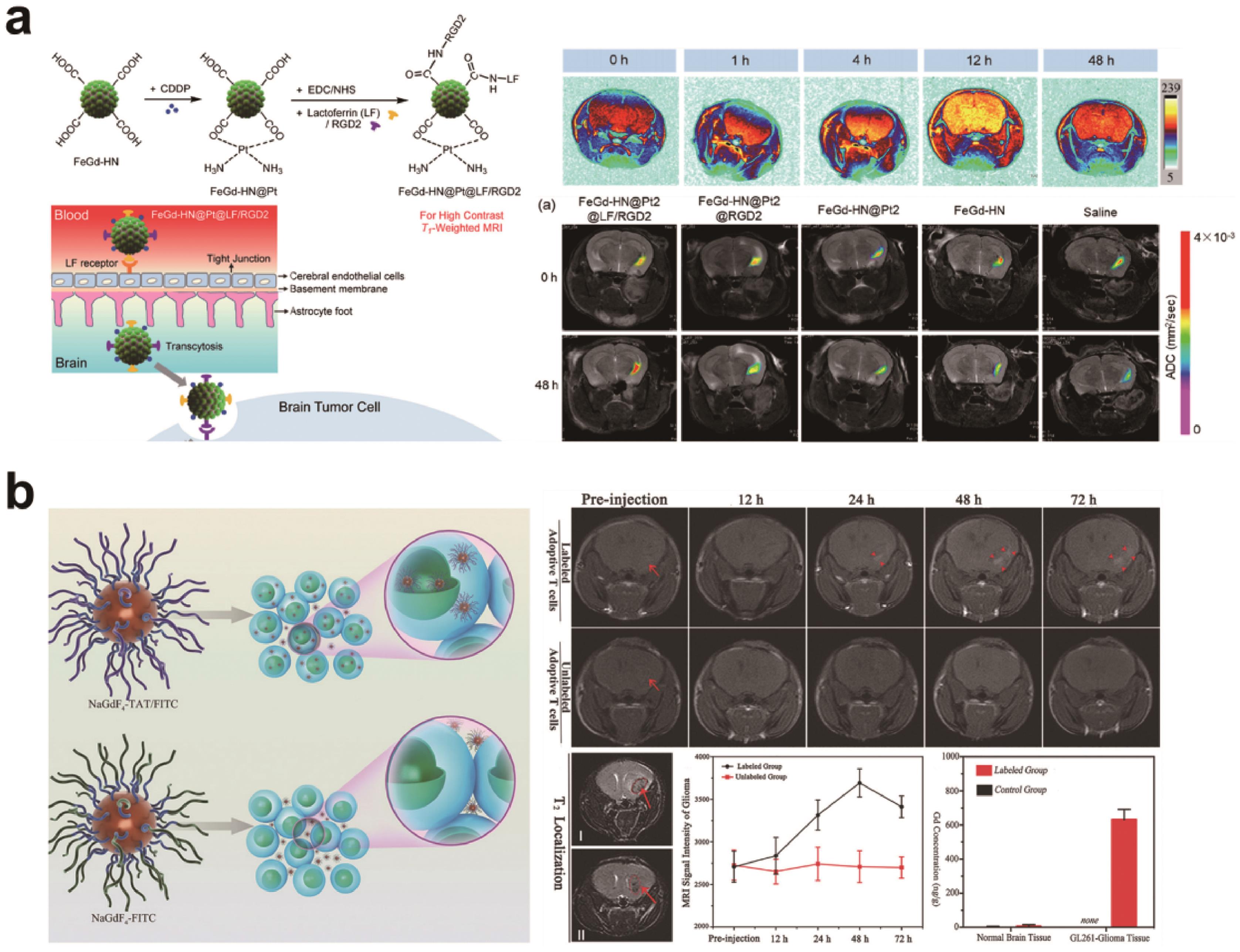
图2 (a) Fe3O4/Gd2O3杂化纳米颗粒的合成以及所合成的RENPs的T1加权磁共振成像效果[36]; (b)细胞穿透性NaGdF4-TAT/FITC和非细胞穿透性NaGdF4-FITC对过继性T细胞标记示意图以及二者在原位胶质瘤中对过继性T细胞的T1加权磁共振成像效果[39]
Fig.2 (a) Schematic illustration for synthesis of the Fe3O4/Gd2O3 hybrid nanoparticles and the results for T1-weighted imaging effect[36]; (b) Schematic illustration for cell-penetrating NaGdF4-TAT/FITC and non-cell-penetrating NaGdF4-FITC labeling of adoptive T cell and T1-weighted magnetic resonance imaging effects of both on adoptive T cell in orthotopic glioma[39]
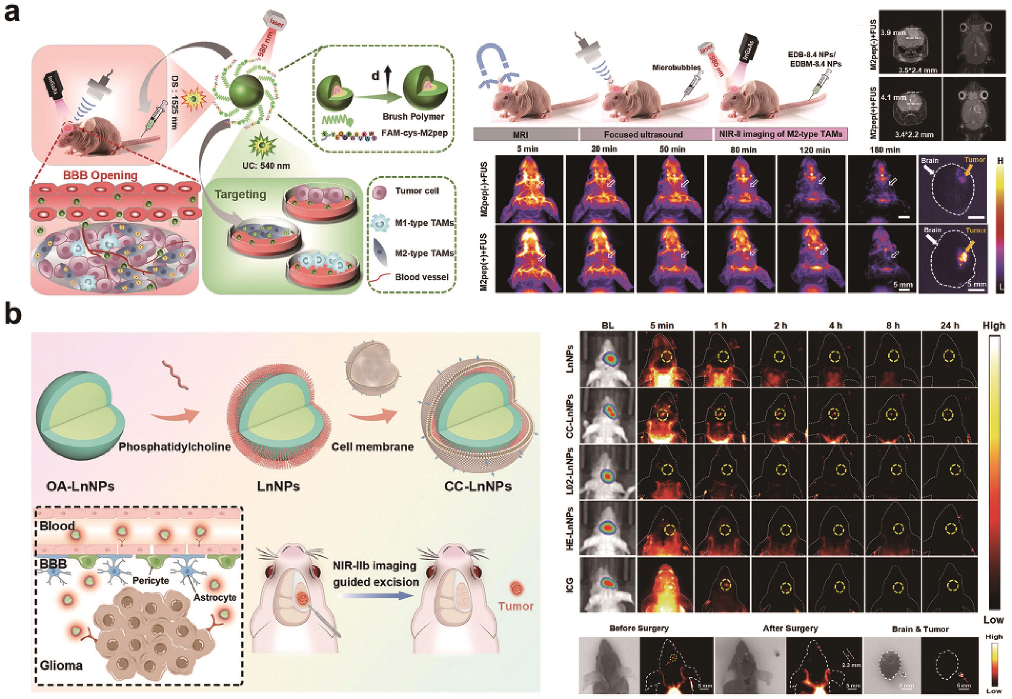
图3 (a) NIR-Ⅱb的EDBM-8.4纳米探针用于体外和体内原位GBM的M2型TAMs靶向成像的示意图以及静脉注射EDB-8.4纳米探针和 EDBM-8.4纳米探针后,在有/无FUS下的小鼠原位GBM的T2加权MR和NIR-Ⅱ荧光成像[45]; (b)脑肿瘤细胞膜包被 RENPs 的制备过程示意图和其在脑肿瘤成像和手术导航中的应用,以及小鼠体内荧光成像和手术的结果[46]
Fig.3 (a) Schematic illustration of NIR-Ⅱb EDBM-8.4 nanoprobes for targeted imaging of M2-type TAMs both in vitro and in orthotopic GBM. And T2-weighted MRI and NIR-Ⅱ fluorescence imaging of orthotopic GBM-bearing mice post intravenous injection of EDB-8.4 NPs and EDBM-8.4 NPs, with or without FUS[45]; (b) Schematic illustration of the preparation processes of brain tumor cell membrane-coated RENPs and its application in brain tumor imaging and surgical navigation. and the results in vivo fluorescence imaging and surgery of mice[46]
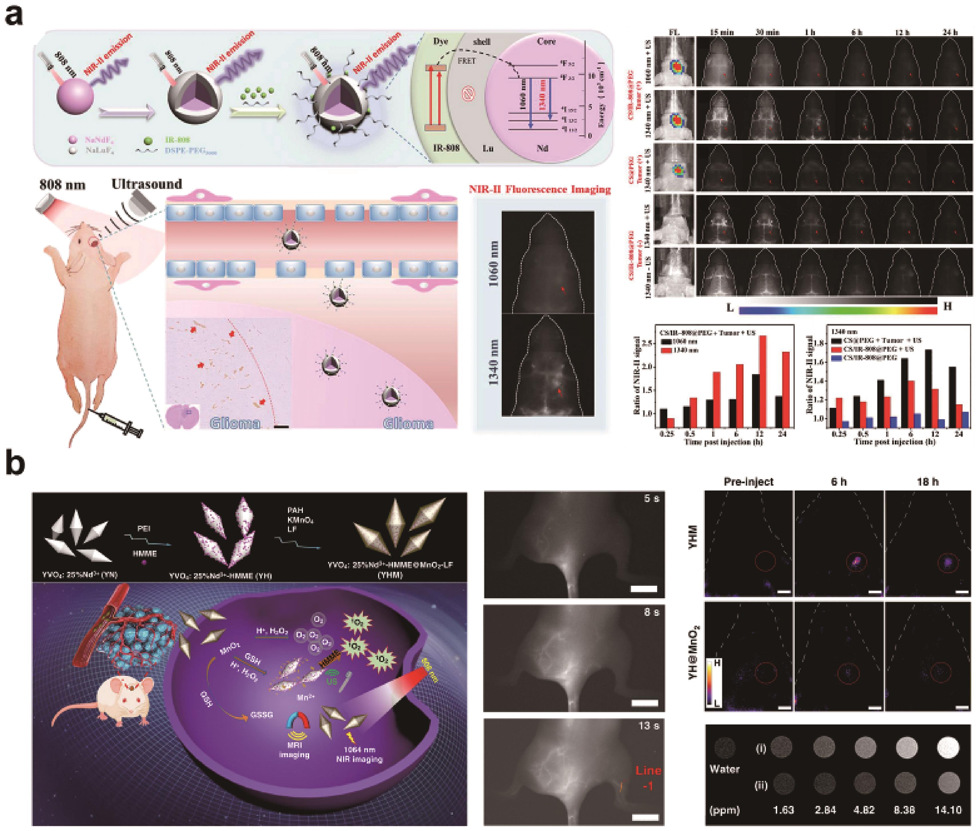
图4 (a) NaNdF4@NaLuF4/IR-808@DSPE-PEG5000 RENPs的设计示意图及其在超声介导的BBB开放条件下对正位胶质母细胞瘤的近红外-Ⅱ荧光成像和超声条件下对正位胶质母细胞瘤在1340 nm发射波长下的活体荧光成像中的应用[52]; (b) YHM的构建、多模态生物成像和治疗作用机制的示意图及其在血管和原位胶质瘤中的NIR-Ⅱ荧光成像效果[53]
Fig.4 (a) Schematic illustration of the design of the NaNdF4@NaLuF4/IR-808@DSPE-PEG5000 RENPs and their applications in NIR-Ⅱ fluorescence imaging of orthotopic glioblastoma under ultrasound-mediated BBB opening and in vivo fluorescence imaging of orthotopic glioblastomas at 1340 nm emission under ultrasound[52]; (b) Schematic diagram of the construction of YHM and main mechanism of multimodal bioimaging and therapy, and its NIR-Ⅱ imaging in vascular and orthotopic gliomas[53]
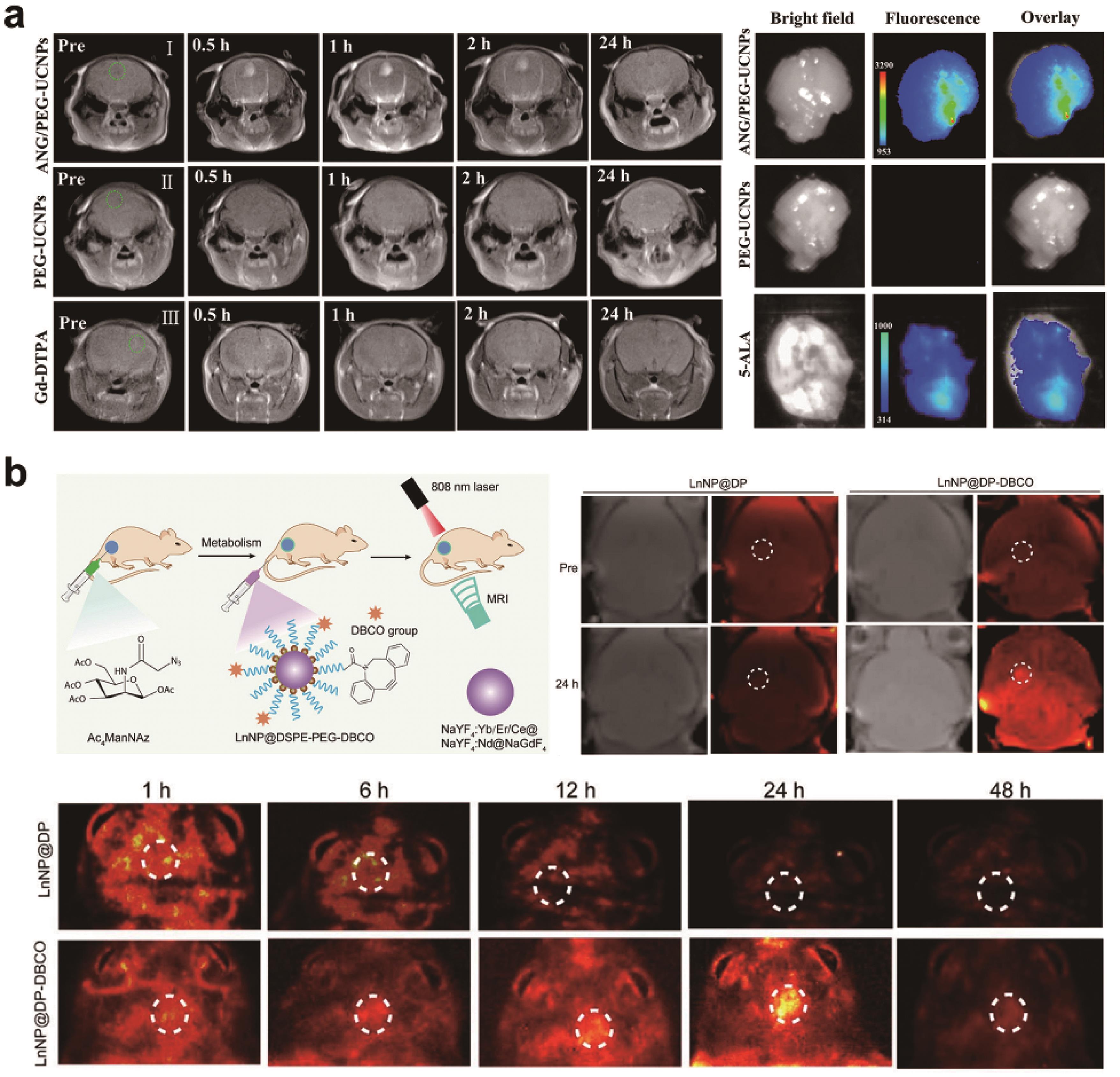
图5 (a)胶质母细胞瘤小鼠在静脉注射ANG/PEG-UCNPs前和注射后不同时间点的体内T1加权磁共振成像,以及胶质母细胞瘤小鼠大脑的体外荧光图像[63]; (b)生物功能化多层纳米颗粒(LnNP@DSPE-PEG-DBCO)用于肿瘤双模态成像的示意图,以及胶质母细胞瘤小鼠在不同时间点的T1加权磁共振成像和NIR-Ⅱb荧光成像评估[65]
Fig.5 (a) In vivo T1-weighted MRI in glioblastoma-bearing mice before and at various time points after the intravenous injections of ANG/PEG-UCNPs, and ex vivo fluorescent images of glioblastoma-bearing brain[63]?; (b) Schematic illustration of biofunctionalization of multilayer nanoparticles (LnNP@DSPE-PEG-DBCO) for dual-modal tumor imaging, and the results of T1-weighted MRI images and NIR-Ⅱb fluorescence imaging in glioblastoma-bearing mice at different time points[65]
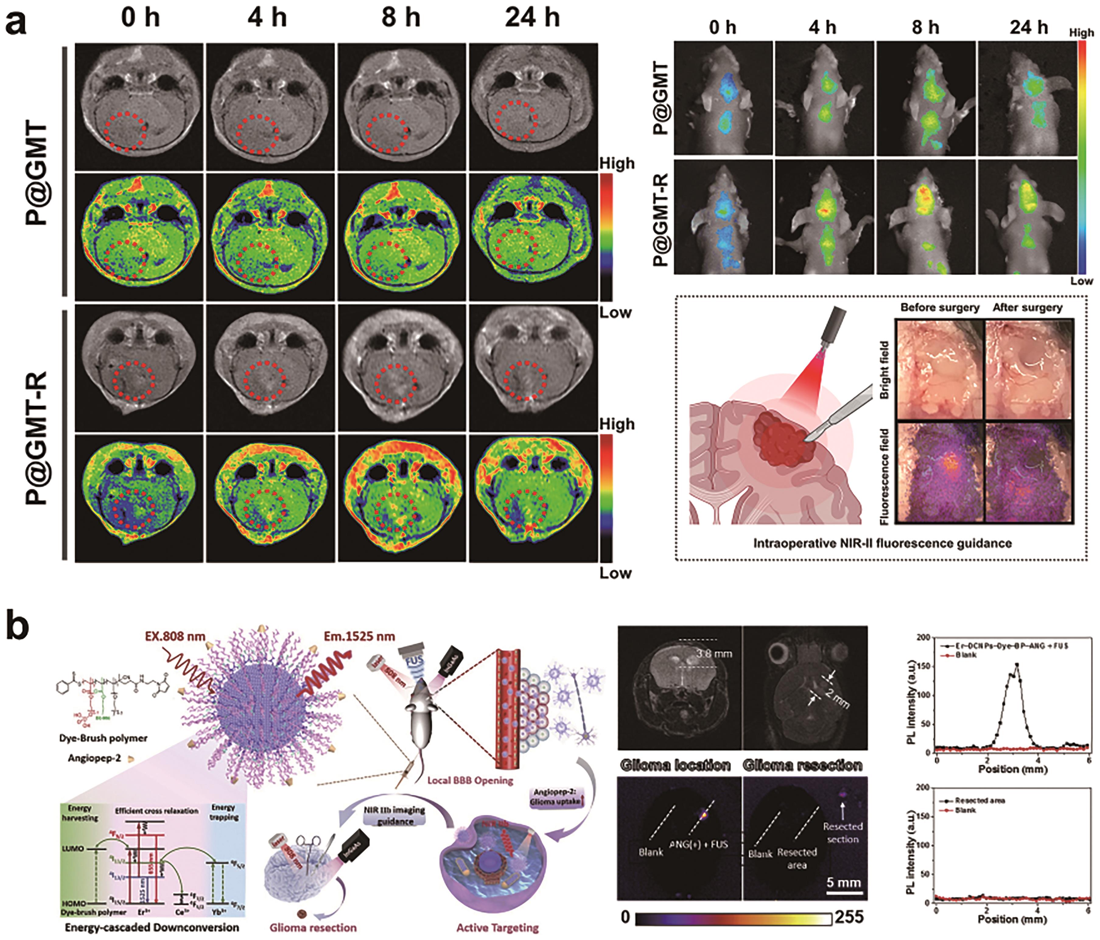
图6 (a) Gd2O3∶Nd3+基纳米材料用于原位胶质瘤的MRI成像以及NIR-Ⅱ荧光成像,并在荧光成像指导下进行手术切除[76]; (b) Er-DCNPs-Dye-BP-ANG的设计、发光机制和生物医学成像应用示意图,以及在NIR-Ⅱb荧光成像指导下进行原位胶质瘤手术切除的评估[50]
Fig.6 (a) Gd2O3∶Nd3+-based nanomaterials for MRI imaging as well as NIR-Ⅱ fluorescence imaging of orthotopic gliomas and fluorescence-guided surgical resection[76]; (b) Schematic illustration of the Er-DCNPs-Dye-BP-ANG design, emission mechanism and the application of biomedical imaging and the assessment about NIR-Ⅱb fluorescence imaging to guide surgical resection of orthotopic gliomas[50]

图7 稀土纳米闪烁体 LaF3∶Ce 的制备示意图,以及在脑肿瘤放射治疗增强作用[85]
Fig.7 Schematic of the preparation of rare-earth nanoscintillator LaF3∶Ce and its role in radiation therapy enhancement of brain tumors[85]
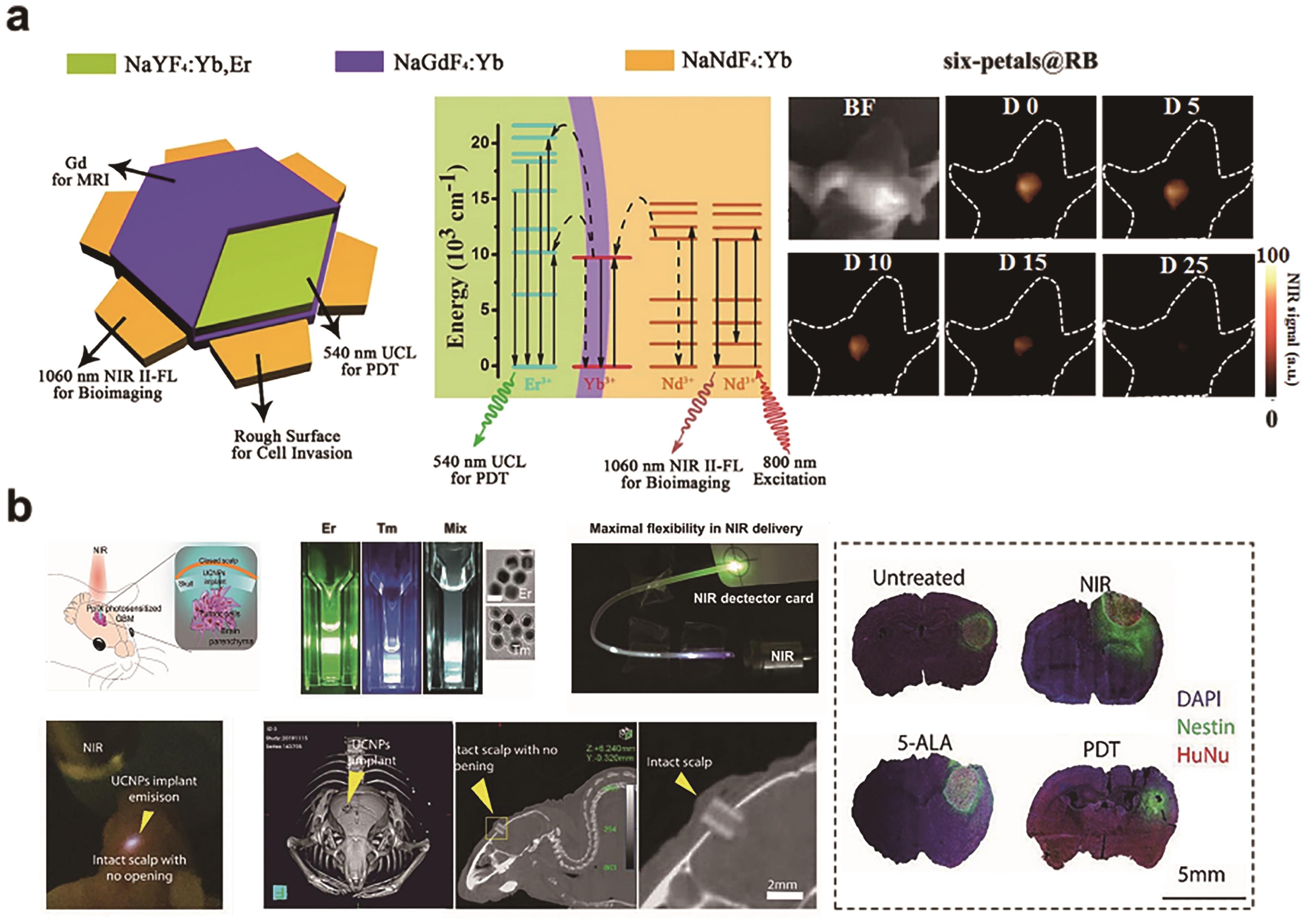
图8 (a)六瓣纳米板的结构示意图和能量传递机制。以及NIR-Ⅱ荧光成像引导six-petals@RB的PDT治疗胶质母细胞瘤小鼠[90]; (b)在小鼠 GBM 模型中植入UCNPs用于无线近红外光导PDT治疗的概念,以及UCNPs植入物的microCT成像结果和PDT的有效性[24]
Fig.8 (a) The structure scheme and energy-transfer mechanisms of the six-petals nanoplates. And NIR-?Ⅱ FL bioimaging guided glioblastoma-bearing mice PDT of six-petals@RB[90]?; (b) The concept of UCNPs implant for wireless NIR-PDT in a mouse GBM model, and the results of microCT scan of UCNPs implant and effectiveness of PDT treatment[24]
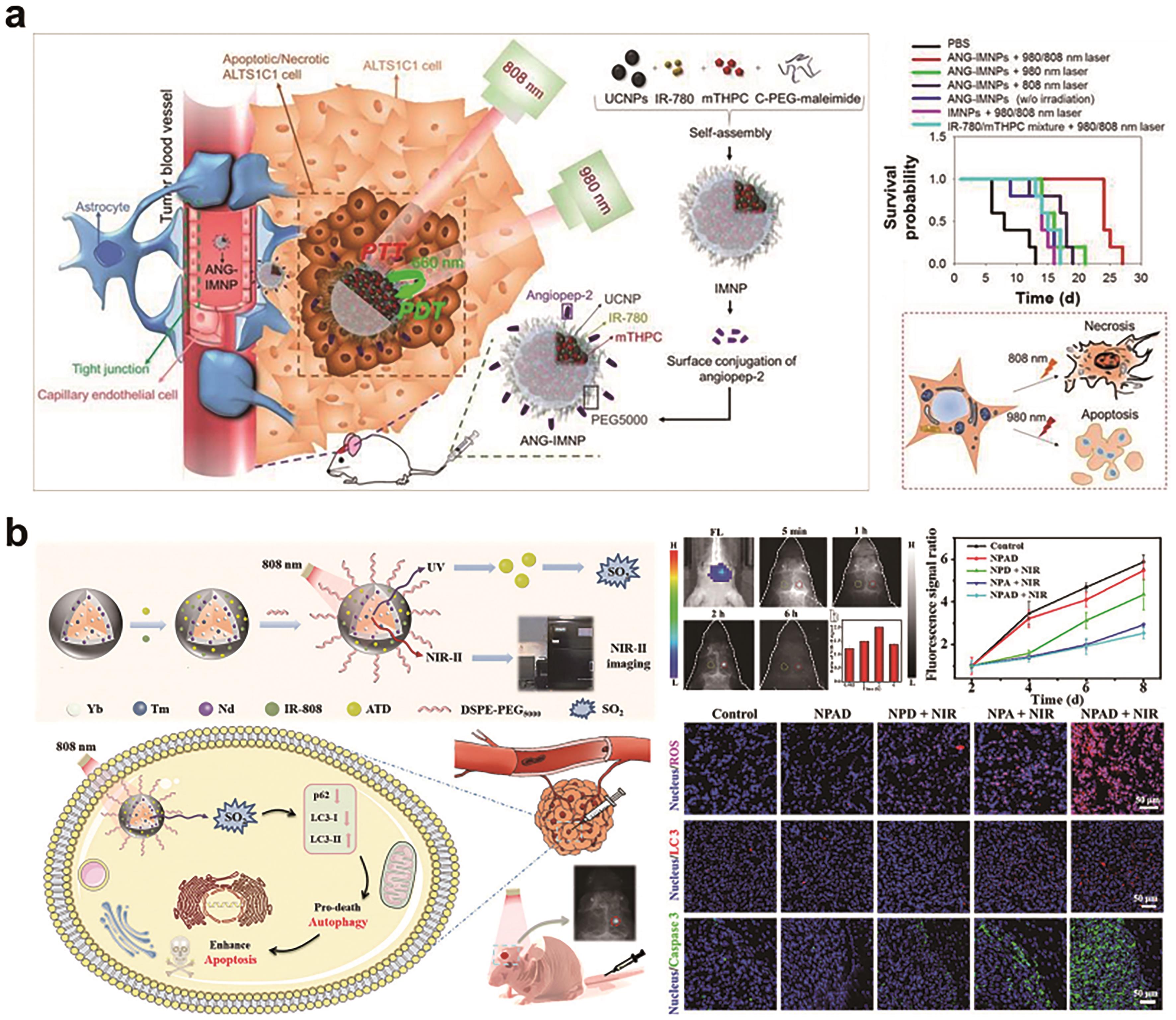
图9 (a) ANG-IMNPs在原位胶质母细胞瘤肿瘤模型中穿过BBB并用于PTT/PDT治疗示意图,以及评估了ANG-IMNPs 的治疗效果[92]; (b) 核壳NaYF4∶Yb/Tm@NaYF4∶Nd@ATD/Dye@DSPE-PEG5000 NPs在原位胶质母细胞瘤的NIR-Ⅱ荧光成像和光控释放二氧化硫治疗中的应用示意图,以及NIR-Ⅱ荧光成像和气体治疗的评估[100]
Fig.9 (a) Illustration of active BBB penetration and the photothermal/photodynamic therapeutic design of ANG-IMNPs in an orthotopic glioblastoma tumor model, and the assessment of ANG-IMNPs treatment effects[92]?; (b) Illustration of the core-shell NaYF4∶?Yb/Tm@NaYF4∶?Nd@ATD/Dye@DSPE-PEG5000 NPs in the NIR-Ⅱ fluorescence imaging of orthotopic glioblastoma and photo-controlled release of SO2 for therapy, and the assessment of NIR-Ⅱ fluorescence imaging and gas therapy[100]
| 1 | SUNG H, FERLAY J, SIEGEL R L, et al. Global cancer statistics 2020: GLOBOCAN estimates of incidence and mortality worldwide for 36 cancers in 185 countries[J]. CA Cancer J Clin, 2021, 71(3): 209-249. |
| 2 | DELGADO-LOPEZ P D, CORRALES-GARCIA E M. Survival in glioblastoma: a review on the impact of treatment modalities[J]. Clin Transl Oncol, 2016, 18(11): 1062-1071. |
| 3 | REIFENBERGER G, WIRSCHING H G, KNOBBE-THOMSEN C B, et al. Advances in the molecular genetics of gliomas-implications for classification and therapy[J]. Nat Rev Clin Oncol, 2017, 14(7): 434-452. |
| 4 | LIN S J, XU H S, ZHANG A K, et al. Prognosis analysis and validation of m6A signature and tumor immune microenvironment in glioma[J]. Front Oncol, 2020, 10: 541401. |
| 5 | SUN C J, HU R Y, LI Z C, et al. An engineered abcb4 expression model reveals the central role of NF-κB in the regulation of drug resistance in zebrafish[J]. Drug Dev Res, 2022, 83(4): 927-939. |
| 6 | WANG Y H, ZHANG F C, XIONG N X, et al. Remodelling and treatment of the blood-brain barrier in glioma[J]. Cancer Manag Res, 2021, 13: 4217-4232. |
| 7 | CAO Y, ZHANG S, LV Z, et al. An intelligent nanoplatform for orthotopic glioblastoma therapy by nonferrous ferroptosis[J]. Adv Funct Mater, 2022, 32(51): 2209227. |
| 8 | YIN N, WANG Y, LIU Y, et al. A cholesterol metabolic regulated hydrogen-bonded organic framework (HOF)-based biotuner for antibody non-dependent immunotherapy tailored for glioblastoma[J]. Adv Mater, 2023: 2303567. |
| 9 | CHARLES N A, HOLLAND E C, GILBERTSON R, et al. The brain tumor microenvironment[J]. Glia, 2012, 60(3): 502-514. |
| 10 | HEGI M E, DISERENS A C, GODARD S, et al. Clinical trial substantiates the predictive value of O-6-methylguanine-DNA methyltransferase promoter methylation in glioblastoma patients treated with temozolomide[J]. Clin Cancer Res, 2004, 10(6): 1871-1874. |
| 11 | OZDEMIR-KAYNAK E, QUTUB A A, YESIL-CELIKTAS O. Advances in glioblastoma multiforme treatment: new models for nanoparticle therapy[J]. Front Physiol, 2018, 9: 170. |
| 12 | GANZ J C. Low grade gliomas[J]. Prog Brain Res, 2022, 268(1): 271-277. |
| 13 | LIU Y, ZHEN W, JIN L, et al. All-in-one theranostic nanoagent with enhanced reactive oxygen species generation and modulating tumor microenvironment ability for effective tumor eradication[J]. ACS Nano, 2018, 12(5): 4886-4893. |
| 14 | LIU Y W, ZHAO K L, REN Y B, et al. Highly plasticized lanthanide luminescence for information storage and encryption applications[J]. Adv Sci, 2022, 9(7): 2105108. |
| 15 | WAN S K, CONG W, SHAO B Q, et al. A library of thermotropic liquid crystals of inorganic nanoparticles and extraordinary performances based on their collective ordering[J]. Nano Today, 2021, 38: 101115. |
| 16 | QU Z B, SHEN J L, LI Q, et al. Near-IR emissive rare-earth nanoparticles for guided surgery[J]. Theranostics, 2020, 10(6): 2631-2644. |
| 17 | ZHAO M N, VAN STRATEN D, BROEKMAN M L D, et al. Nanocarrier-based drug combination therapy for glioblastoma[J]. Theranostics, 2020, 10(3): 1355-1372. |
| 18 | LIU Y L, AI K L, LIU J H, et al. A high-performance ytterbium-based nanoparticulate contrast agent for in vivo X-ray computed tomography imaging[J]. Angew Chem Int Ed, 2012, 51(6): 1437-1442. |
| 19 | VISWANATHAN S, KOVACS Z, GREEN K N, et al. Alternatives to gadolinium-based metal chelates for magnetic resonance imaging[J]. Chem Rev, 2010, 110(5): 2960-3018. |
| 20 | CAO Y, XU L J, KUANG Y, et al. Gadolinium-based nanoscale MRI contrast agents for tumor imaging[J]. J Mater Chem B, 2017, 5(19): 3431-3461. |
| 21 | ZHONG Y, DAI H. A mini-review on rare-earth down-conversion nanoparticles for NIR-Ⅱ imaging of biological systems[J]. Nano Res, 2020, 13(5): 1281-1294. |
| 22 | ESCUDERO A, BECERRO A I, CARRILLO-CARRION C, et al. Rare earth based nanostructured materials: synthesis, functionalization, properties and bioimaging and biosensing applications[J]. Nanophotonics, 2017, 6(5): 881-921. |
| 23 | LE DUC G, MILADI I, ALRIC C, et al. Toward an image-guided microbeam radiation therapy using gadolinium-based nanoparticles[J]. ACS Nano, 2011, 5(12): 9566-9574. |
| 24 | TEH D B L, BANSAL A, CHAI C, et al. A flexi-PEGDA upconversion implant for wireless brain photodynamic therapy[J]. Adv Mater, 2020, 32(29): 2001459. |
| 25 | GAI S L, LI C X, YANG P P, et al. Recent progress in rare earth micro/nanocrystals: soft chemical synthesis, luminescent properties, and biomedical applications [J]. Chem Rev, 2014, 114(4): 2343-2389. |
| 26 | AVRAM D, COJOCARU B, URDA A, et al. Pure and almost pure NIR emission of Tm and Tm,Yb-CeO2 under UV, X-ray and NIR up-conversion excitation: key roles of level selective antenna sensitization and charge-compensation[J]. Phys Chem Chem Phys, 2015, 17(46): 30988-30992. |
| 27 | NOREK M, PETERS J A. MRI contrast agents based on dysprosium or holmium[J]. Prog Nucl Magn Reson Spectrosc, 2011, 59(1): 64-82. |
| 28 | FRIES P H, BELORIZKY E. Electronic relaxation of paramagnetic metal ions and NMR relaxivity in solution: critical analysis of various approaches and application to a Gd(Ⅲ)-based contrast agent[J]. J Chem Phys, 2005, 123(12): 124510. |
| 29 | CHEN X F, SONG J B, CHEN X Y, et al. X-ray-activated nanosystems for theranostic applications[J]. Chem Soc Rev, 2019, 48(11): 3073-3101. |
| 30 | LUSIC H, GRINSTAFF M W. X-ray-computed tomography contrast agents[J]. Chem Rev, 2013, 113(3): 1641-1666. |
| 31 | MCDONALD M A, WATKIN K L. Small particulate gadolinium oxide and gadolinium oxide albumin microspheres as multimodal contrast and therapeutic agents[J]. Invest Radiol, 2003, 38(6): 305-310. |
| 32 | PARK J Y, BAEK M J, CHOI E S, et al. Paramagnetic ultrasmall gadolinium oxide nanoparticles as advanced T1 MRI contrast agent: account for large longitudinal relaxivity, optimal particle diameter, and in vivo T1 MR images[J]. ACS Nano, 2009, 3(11): 3663-3669. |
| 33 | FAUCHER L, TREMBLAY M, LAGUEUX J, et al. Rapid synthesis of pegylated ultrasmall gadolinium oxide nanoparticles for cell labeling and tracking with MRI[J]. ACS Appl Mater Interfaces, 2012, 4(9): 4506-4515. |
| 34 | GU W, SONG G, LI S, et al. Chlorotoxin-conjugated, PEGylated Gd2O3 nanoparticles as a glioma-specific magnetic resonance imaging contrast agent[J]. RSC Adv, 2014, 4(91): 50254-50260. |
| 35 | LI W, QIU J H, LI X L, et al. BBB pathophysiology-independent delivery of siRNA in traumatic brain injury[J]. Sci Adv, 2021, 7(1): eabd6889. |
| 36 | SHEN Z, LIU T, LI Y, et al. Fenton-reaction-acceleratable magnetic nanoparticles for ferroptosis therapy of orthotopic brain tumors[J]. ACS Nano, 2018, 12(11): 11355-11365. |
| 37 | SHAO C, LI S, GU W, et al. Multifunctional gadolinium-doped manganese carbonate nanoparticles for targeted MR/fluorescence imaging of tiny brain gliomas[J]. Anal Chem, 2015, 87(12): 6251-6257. |
| 38 | LI T, MURPHY S, KISELEV B, et al. A new interleukin-13 amino-coated gadolinium metallofullerene nanoparticle for targeted MRI detection of glioblastoma tumor cells[J]. J Am Chem Soc, 2015, 137(24): 7881-7888. |
| 39 | ZHANG H, WU Y, WANG J, et al. In vivo MR imaging of glioma recruitment of adoptive T-cells labeled with NaGdF4-TAT nanoprobes[J]. Small, 2018, 14(3): 1702951. |
| 40 | KENRY, DUAN Y, LIU B. Recent advances of optical imaging in the second near-infrared window[J]. Adv Mater, 2018, 30(47): 1802394. |
| 41 | CHEN S Y, MIAO H, JIANG X Y, et al. Starlike polymer brush-based ultrasmall nanoparticles with simultaneously improved NIR-Ⅱ fluorescence and blood circulation for efficient orthotopic glioblastoma imaging[J]. Biomaterials, 2021, 275: 120916. |
| 42 | ZHONG Y, MA Z, ZHU S, et al. Boosting the down-shifting luminescence of rare-earth nanocrystals for biological imaging beyond 1500 nm[J]. Nat Commun, 2017, 8: 737. |
| 43 | LV Z J, JIN L H, GAO W H, et al. Novel YOF-based theranostic agents with a cascade effect for NIR-Ⅱ fluorescence imaging and synergistic starvation/photodynamic therapy of orthotopic gliomas[J]. ACS Appl Mater Interfaces, 2022, 14(27): 30523-30532. |
| 44 | FAN Y, ZHANG F. A new generation of NIR-Ⅱ probes: lanthanide-based nanocrystals for bioimaging and biosensing[J]. Adv Opt Mater, 2019, 7(7): 1801417. |
| 45 | ZHU H, REN F, WANG T, et al. Targeted immunoimaging of tumor-associated macrophages in orthotopic glioblastoma by the NIR-Ⅱb nanoprobes[J]. Small, 2022, 18(30): 2202201. |
| 46 | WANG Z J, ZHANG M, CHI S Y, et al. Brain tumor cell membrane-coated lanthanide-doped nanoparticles for NIR-Ⅱb luminescence imaging and surgical navigation of glioma[J]. Adv Healthc Mater, 2022, 11(16): 2200521. |
| 47 | ZHANG X B, CHEN W W, XIE X Y, et al. Boosting luminance energy transfer efficiency in upconversion nanoparticles with an energy-concentrating zone[J]. Angew Chem Int Ed, 2019, 58(35): 12117-12122. |
| 48 | XUE B, WANG D, TU L P, et al. Ultrastrong absorption meets ultraweak absorption: unraveling the energy-dissipative routes for dye-sensitized upconversion luminescence[J]. J Phys Chem Lett, 2018, 9(16): 4625-4631 . |
| 49 | ZOU W Q, VISSER C, MADURO J A, et al. Broadband dye-sensitized upconversion of near-infrared light[J]. Nat Photonics, 2012, 6(8): 560-564. |
| 50 | REN F, LIU H, ZHANG H, et al. Engineering NIR-Ⅱb fluorescence of Er-based lanthanide nanoparticles for through-skull targeted imaging and imaging-guided surgery of orthotopic glioma[J]. Nano Today, 2020, 34: 100905. |
| 51 | PENG J J, XU W, TEOH C L, et al. High-efficiency in vitro and in vivo detection of Zn2+-dye-assembled upconversion nanoparticles[J]. J Am Chem Soc, 2015, 137(6): 2336-2342. |
| 52 | LIU Z, REN F, ZHANG H, et al. Boosting often overlooked long wavelength emissions of rare-earth nanoparticles for NIR-Ⅱ fluorescence imaging of orthotopic glioblastoma[J]. Biomaterials, 2019, 219: 119364. |
| 53 | LV Z, JIN L, CAO Y, et al. A nanotheranostic agent based on Nd3+-doped YVO4 with blood-brain-barrier permeability for NIR-Ⅱ fluorescence imaging/magnetic resonance imaging and boosted sonodynamic therapy of orthotopic glioma[J]. Light Sci Appl, 2022, 11(1): 116. |
| 54 | PAN D, ROESSL E, SCHLOMKA J P, et al. Computed tomography in color: nanoK-enhanced spectral CT molecular imaging[J]. Angew Chem Int Ed, 2010, 49(50): 9635-9639. |
| 55 | JIN X, FANG F, LIU J, et al. An ultrasmall and metabolizable PEGylated NaGdF4∶Dy nanoprobe for high-performance T1/T2-weighted MR and CT multimodal imaging[J]. Nanoscale, 2015, 7(38): 15680-15688. |
| 56 | HU Y, ZHOU Y Q, ZHAO N N, et al. Multifunctional pDNA-conjugated polycationic Au nanorod-coated Fe3O4 hierarchical nanocomposites for trimodal imaging and combined photothermal/gene therapy[J]. Small, 2016, 12(18): 2459-2468. |
| 57 | LIU Z, LI Z, LIU J, et al. Long-circulating Er3+-doped Yb2O3 up-conversion nanoparticle as an in vivo X-Ray CT imaging contrast agent[J]. Biomaterials, 2012, 33(28): 6748-6757. |
| 58 | NI D L, BU W B, ZHANG S J, et al. Single Ho3+-doped upconversion nanoparticles for high-performance T2-weighted brain tumor diagnosis and MR/UCL/CT multimodal imaging[J]. Adv Funct Mater, 2014, 24(42): 6613-6620. |
| 59 | LEE D E, KOO H, SUN I C, et al. Multifunctional nanoparticles for multimodal imaging and theragnosis[J]. Chem Soc Rev, 2012, 41(7): 2656-2672. |
| 60 | XUE D, LIU Y, JIN L, et al. Novel multifunctional theranostic nanoagents based on Ho3+ for CT/MRI dual-modality imaging-guided photothermal therapy[J]. Sci China Chem, 2021, 64(4): 558-564. |
| 61 | BOTTRILL M, NICHOLAS L K, LONG N J. Lanthanides in magnetic resonance imaging[J]. Chem Soc Rev, 2006, 35(6): 557-571. |
| 62 | JIN J, XU Z, ZHANG Y, et al. Upconversion nanoparticles conjugated with Gd3+-DOTA and RGD for targeted dual-modality imaging of brain tumor xenografts[J]. Adv Healthc Mater, 2013, 2(11): 1501-1512. |
| 63 | NI D, ZHANG J, BU W, et al. Dual-targeting upconversion nanoprobes across the blood-brain barrier for magnetic resonance/fluorescence imaging of intracranial glioblastoma[J]. ACS Nano, 2014, 8(2): 1231-1242. |
| 64 | LIU Y, LI L, GUO Q, et al. Novel Cs-based upconversion nanoparticles as dual-modal CT and UCL imaging agents for chemo-photothermal synergistic therapy[J]. Theranostics, 2016, 6(10): 1491-1505. |
| 65 | LUO Z, HU D, GAO D, et al. High-specificity in vivo tumor imaging using bioorthogonal NIR-Ⅱb nanoparticles[J]. Adv Mater, 2021, 33(49): 2102950. |
| 66 | DE BOER E, HARLAAR N J, TARUTTIS A, et al. Optical innovations in surgery[J]. Br J Surg, 2015, 102(2): E56-E72. |
| 67 | DEBIE P, HERNOT S. Emerging fluorescent molecular tracers to guide intra-operative surgical decision-making[J]. Front Pharmacol, 2019, 10: 510. |
| 68 | YANG L L, SAJJA H K, CAO Z H, et al. uPAR-targeted optical imaging contrasts as theranostic agents for tumor margin detection[J]. Theranostics, 2014, 4(1): 106-118. |
| 69 | KUHNT D, BECKER A, GANSLANDT O, et al. Correlation of the extent of tumor volume resection and patient survival in surgery of glioblastoma multiforme with high-field intraoperative MRI guidance[J]. Neuro-Oncology, 2011, 13(12): 1339-1348. |
| 70 | CHI C, DU Y, YE J, et al. Intraoperative imaging-guided cancer surgery: from current fluorescence molecular imaging methods to future multi-modality imaging technology[J]. Theranostics, 2014, 4(11): 1072-1084. |
| 71 | GU K, XU Y, LI H, et al. Real-time tracking and in vivo visualization of β-galactosidase activity in colorectal tumor with a ratiometric near-infrared fluorescent probe[J]. J Am Chem Soc, 2016, 138(16): 5334-5340. |
| 72 | WANG C, FAN W, ZHANG Z, et al. Advanced nanotechnology leading the way to multimodal imaging-guided precision surgical therapy[J]. Adv Mater, 2019, 31(49): 1904329. |
| 73 | VAHRMEIJER A L, HUTTEMAN M, VAN DER VORST J R, et al. Image-guided cancer surgery using near-infrared fluorescence[J]. Nat Rev Clin Oncol, 2013, 10(9): 507-518. |
| 74 | HONG G, ANTARIS A L, DAI H. Near-infrared fluorophores for biomedical imaging[J]. Nat Biomed Eng, 2017, 1(1): 0010. |
| 75 | HE S, SONG J, QU J, et al. Crucial breakthrough of second near-infrared biological window fluorophores: design and synthesis toward multimodal imaging and theranostics[J]. Chem Soc Rev, 2018, 47(12): 4258-4278. |
| 76 | YIN N, WANG Y, HUANG Y, et al. A biodegradable nanocapsule for through-skull NIR-Ⅱ fluorescence imaging/magnetic resonance imaging and selectively enhanced radio-chemotherapy for orthotopic glioma[J]. Nano Today, 2022, 46: 101619. |
| 77 | ZHANG H, WANG T T, QIU W B, et al. Monitoring the opening and recovery of the blood-brain barrier with noninvasive molecular imaging by biodegradable ultrasmall Cu2- xSe nanoparticles[J]. Nano Lett, 2018, 18(8): 4985-4992. |
| 78 | DELANEY G, JACOB S, FEATHERSTONE C, et al. The role of radiotherapy in cancer treatment-estimating optimal utilization from a review of evidence-based clinical guidelines[J]. Cancer, 2005, 104(6): 1129-1137. |
| 79 | LIU Y, ZHEN W, WANG Y, et al. Na2S2O8 nanoparticles trigger antitumor immunotherapy through reactive oxygen species storm and surge of tumor osmolarity[J]. J Am Chem Soc, 2020, 142(52): 21751-21757. |
| 80 | MISAWA M, TAKAHASHI J. Generation of reactive oxygen species induced by gold nanoparticles under X-ray and UV irradiations[J]. Nanomedicine, 2011, 7(5): 604-614. |
| 81 | KOBAYASHI K, USAMI N, PORCEL E, et al. Enhancement of radiation effect by heavy elements[J]. Mutat Res Rev Mutat Res, 2010, 704(1/2/3): 123-131. |
| 82 | MOWAT P, MIGNOT A, RIMA W, et al. In vitro radiosensitizing effects of ultrasmall gadolinium based particles on tumour cells[J]. J Nanosci Nanotechnol, 2011, 11(9): 7833-7839. |
| 83 | LE DUC G, ROUX S, PARUTA-TUAREZ A, et al. Advantages of gadolinium based ultrasmall nanoparticles vs molecular gadolinium chelates for radiotherapy guided by MRI for glioma treatment[J]. Cancer Nanotechnol, 2014, 5(1): 4. |
| 84 | BRIGGS A, CORDE S, OKTARIA S, et al. Cerium oxide nanoparticles: influence of the high-Z component revealed on radioresistant 9L cell survival under X-ray irradiation[J]. Nanomedicine, 2013, 9(7): 1098-1105. |
| 85 | BULIN A L, BROEKGAARDEN M, CHAPUT F, et al. Radiation dose-enhancement is a potent radiotherapeutic effect of rare-earth composite nanoscintillators in preclinical models of glioblastoma[J]. Adv Sci, 2020, 7(20): 2001675. |
| 86 | YANG Z, FAN W, ZOU J, et al. Precision cancer theranostic platform by in situ polymerization in perylene diimide-hybridized hollow mesoporous organosilica nanoparticles[J]. J Am Chem Soc, 2019, 141(37): 14687-14698. |
| 87 | SUN W, LUO L, FENG Y, et al. Aggregation-induced emission gold clustoluminogens for enhanced low-dose X-ray-induced photodynamic therapy[J]. Angew Chem Int Ed, 2020, 59(25): 9914-9921. |
| 88 | LV Z, CAO Y, XUE D, et al. A multiphoton transition activated iron based metal organic framework for synergistic therapy of photodynamic therapy/chemodynamic therapy/chemotherapy for orthotopic gliomas[J]. J Mater Chem B, 2023, 11(5): 1100-1107. |
| 89 | PARK Y, KIM H M, KIM J H, et al. Theranostic probe based on lanthanide-doped nanoparticles for simultaneous in vivo dual-modal imaging and photodynamic therapy[J]. Adv Mater, 2012, 24(42): 5755-5761. |
| 90 | WANG P, WANG C, LU L, et al. Kinetics-mediate fabrication of multi-model bioimaging lanthanide nanoplates with controllable surface roughness for blood brain barrier transportation[J]. Biomaterials, 2017, 141: 223-232. |
| 91 | ZHANG M, WANG W, MOHAMMADNIAEI M, et al. Upregulating aggregation-induced-emission nanoparticles with blood-tumor-barrier permeability for precise photothermal eradication of brain tumors and induction of local immune responses[J]. Adv Mater, 2021, 33(22): 2008802. |
| 92 | TSAI Y C, VIJAYARAGHAVAN P, CHIANG W H, et al. Targeted delivery of functionalized upconversion nanoparticles for externally triggered photothermal/photodynamic therapies of brain glioblastoma[J]. Theranostics, 2018, 8(5): 1435-1448. |
| 93 | YIN N, WANG Y, HUANG Y, et al. Modulating nanozyme-based nanomachines via microenvironmental feedback for differential photothermal therapy of orthotopic gliomas[J]. Adv Sci, 2023, 10(3): 2204937. |
| 94 | FAN W, BU W, ZHANG Z, et al. X-ray radiation-controlled NO-release for on-demand depth-independent hypoxic radiosensitization[J]. Angew Chem Int Ed, 2015, 54(47): 14026-14030. |
| 95 | QIANGLAN L, TONG L, MIN X, et al. SO2 prodrug doped nanorattles with extra-high drug payload for ldquocollusion inside and outsiderdquo photothermal/pH triggered-gas therapy[J]. Biomaterials, 2020, 257: 120236. |
| 96 | SUN R, LIU X C, LI G Z, et al. Photoactivated H2 nanogenerator for enhanced chemotherapy of bladder cancer[J]. ACS Nano, 2020, 14(7): 8135-8148. |
| 97 | CHUNG M F, LIU H Y, LIN K J, et al. A pH-responsive carrier system that generates NO bubbles to trigger drug release and reverse P-glycoprotein-mediated multidrug resistance[J]. Angew Chem Int Ed, 2015, 54(34): 9890- 9893. |
| 98 | DONG H, DU S R, ZHENG X Y, et al. Lanthanide nanoparticles: from design toward bioimaging and therapy[J]. Chem Rev, 2015, 115(19): 10725-10815. |
| 99 | NACZYNSKI D J, TAN M C, ZEVON M, et al. Rare-earth-doped biological composites as in vivo shortwave infrared reporters[J]. Nat Commun, 2013, 4: 2199. |
| 100 | LIU Z, YUN B, HAN Y, et al. Dye-sensitized rare earth nanoparticles with up/down conversion luminescence for on-demand gas therapy of glioblastoma guided by NIR-Ⅱ fluorescence imaging[J]. Adv Healthc Mater., 2022, 11(3): 2102042. |
| 101 | YUN B F, GU Z P, LIU Z, et al. Reducing chemo-/radioresistance to boost the therapeutic efficacy against temozolomide-resistant glioblastoma[J]. ACS Appl Mater Interfaces, 2022, 14(34): 38617-38630. |
| [1] | 赵欣雨, 秦作佳, 张晓兵, 袁林. 近红外二区激活型小分子荧光探针研究进展[J]. 应用化学, 2024, 41(1): 39-59. |
| [2] | 王超宇, 赵璐, 王科伟, 白云峰, 冯锋. 共价有机框架的构筑策略及其在肿瘤治疗中应用的研究进展[J]. 应用化学, 2023, 40(7): 976-994. |
| [3] | 卜芃, 李宏亮. 稀土Yb3+/Tm3+掺杂NaGd(MO4)2荧光粉的制备及其光致发光[J]. 应用化学, 2023, 40(3): 374-379. |
| [4] | 王俊荣, 孙倩倩, 朱国庆, 钱彦荣, 李春霞. 稀土掺杂正交发光纳米晶: 从基础到前沿应用[J]. 应用化学, 2023, 40(11): 1475-1493. |
| [5] | 杜慧, 姚晨阳, 彭皓, 姜波, 李顺祥, 姚俊烈, 郑方, 杨方, 吴爱国. 过渡金属掺杂磁性纳米粒子在生物医学领域中的研究进展[J]. 应用化学, 2022, 39(3): 391-406. |
| [6] | 郝斌,赵文武,郁建元,刘进强,刘剑,董秀珍,王秀文. 荧光粉Ba5-3x/2B4O11∶xEu3+的制备及发光性能[J]. 应用化学, 2019, 36(5): 548-553. |
| [7] | 王涛,马拉毛草,马恒昌. 基于聚集诱导发光荧光探针的细胞成像研究进展[J]. 应用化学, 2018, 35(10): 1155-1165. |
| [8] | 黄子珂, 刘超, 付强强, 李进, 邹建梅, 谢斯滔, 邱丽萍. 核酸适配体荧光探针在生化分析和生物成像中的研究进展[J]. 应用化学, 2018, 35(1): 28-39. |
| [9] | 周佳, 倪赟, 张承武, 仇兴汉, 赵燕菲, 白磊, 张高宾, 李林. 嘧啶基双光子荧光染料的设计合成与生物成像[J]. 应用化学, 2017, 34(12): 1450-1456. |
| [10] | 周佳, 倪赟, 张承武, 仇兴汉, 赵燕菲, 白磊, 张高宾, 李林. 嘧啶基双光子荧光染料的设计合成与生物成像[J]. 应用化学, 2017, 34(12): 0-0. |
| [11] | 李海东,姚起超,樊江莉,杜健军,彭孝军. 钯离子荧光探针的研究进展[J]. 应用化学, 2016, 33(10): 1099-1114. |
| 阅读次数 | ||||||
|
全文 |
|
|||||
|
摘要 |
|
|||||