应用化学 ›› 2022, Vol. 39 ›› Issue (11): 1726-1734.DOI: 10.19894/j.issn.1000-0518.220041

• 研究论文 • 上一篇    下一篇

高荧光量子产率钕、氮双掺杂碳点用于柳氮磺吡啶检测及Hela细胞成像

宋金萍1, 马琦1(), 梁晓敏2, 尚建鹏1, 董川()   

  1. 1.山西大同大学化学与化工学院,应用化学研究所,大同 037009
    2.山西师范大学化学与材料科学学院,临汾 041004) 3. (山西大学环境科学研究所,太原 030006
  • 收稿日期:2022-02-21 接受日期:2022-04-20 出版日期:2022-11-01 发布日期:2022-11-09
  • 通讯作者: 马琦,董川
  • 基金资助:
    山西省自然科学基金(201901D111309);大同市工业重点研发计划项目(2019027)

Neodymium and Nitrogen Co‑doped Carbon Dots with High Fluorescence Quantum Yield for Detection of Sulfasalazine and Hela Cell Imaging

Jin-Ping SONG1, Qi MA1(), Xiao-Min LIANG2, Jian-Peng SHANG1, Chuan DONG()   

  1. 1.College of Chemistry and Chemical Engineering,Institute of Applied Chemistry,Shanxi Datong University,Datong 037009,China
    2.School of Chemistry and Material Science,Shanxi Normal University,Linfen 041004,China
    3.Institute for Environmental Science,Shanxi University,Taiyuan 030006,China
  • Received:2022-02-21 Accepted:2022-04-20 Published:2022-11-01 Online:2022-11-09
  • Contact: Qi MA,Chuan DONG
  • About author:dc@sxu.edu.cn
    maqihx@163.com
  • Supported by:
    the Natural Science Foundation of Shanxi Province(201901D111309);the Research Project Foundation of Datong City(2019027)

摘要:

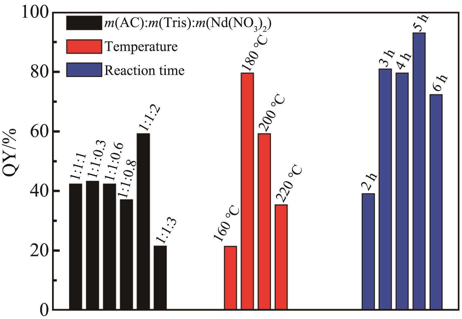
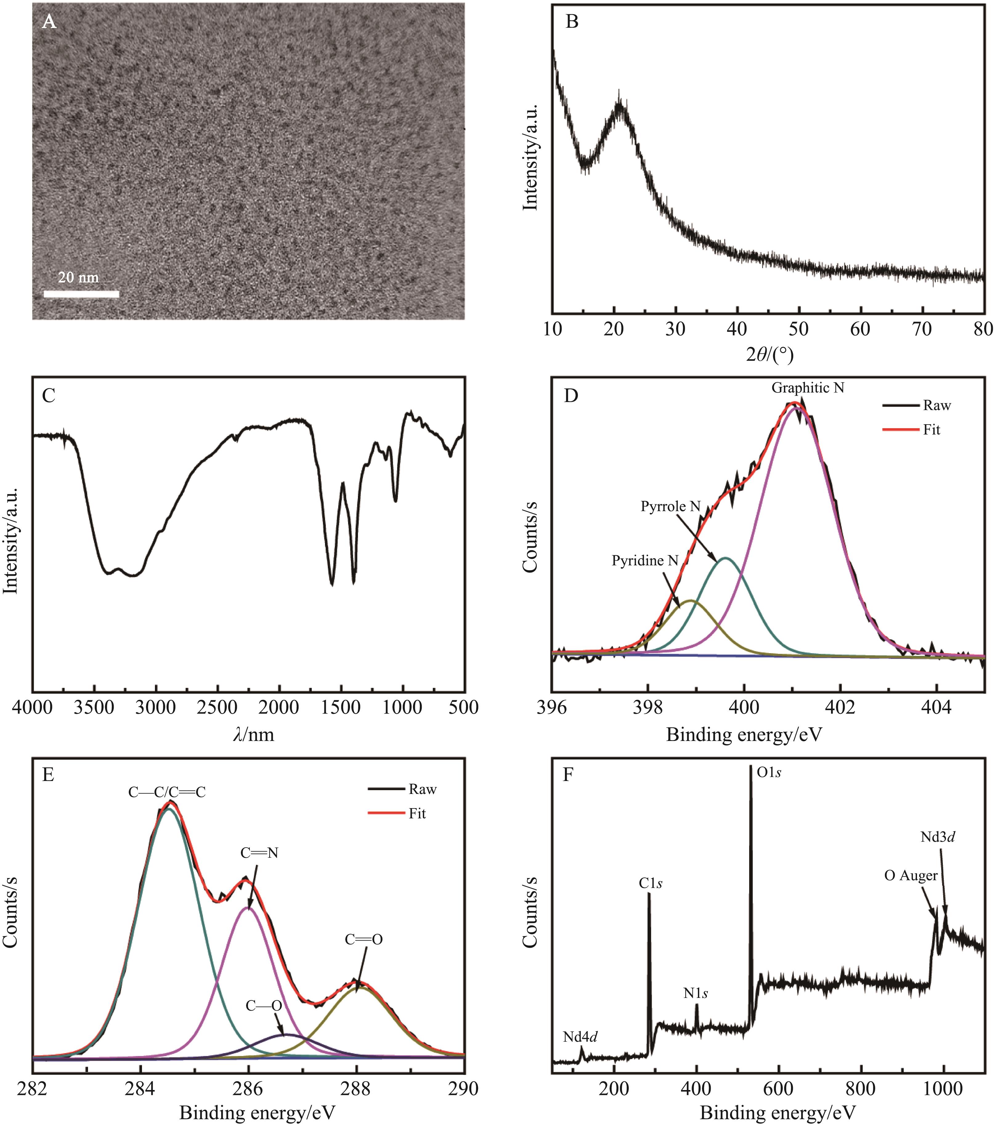
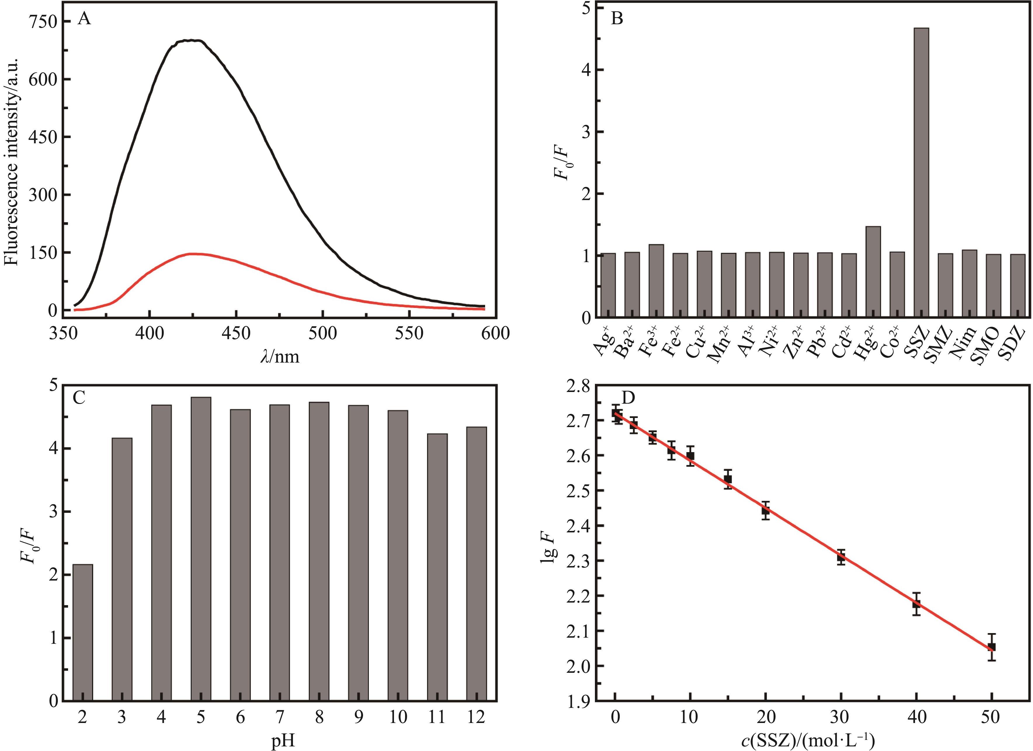
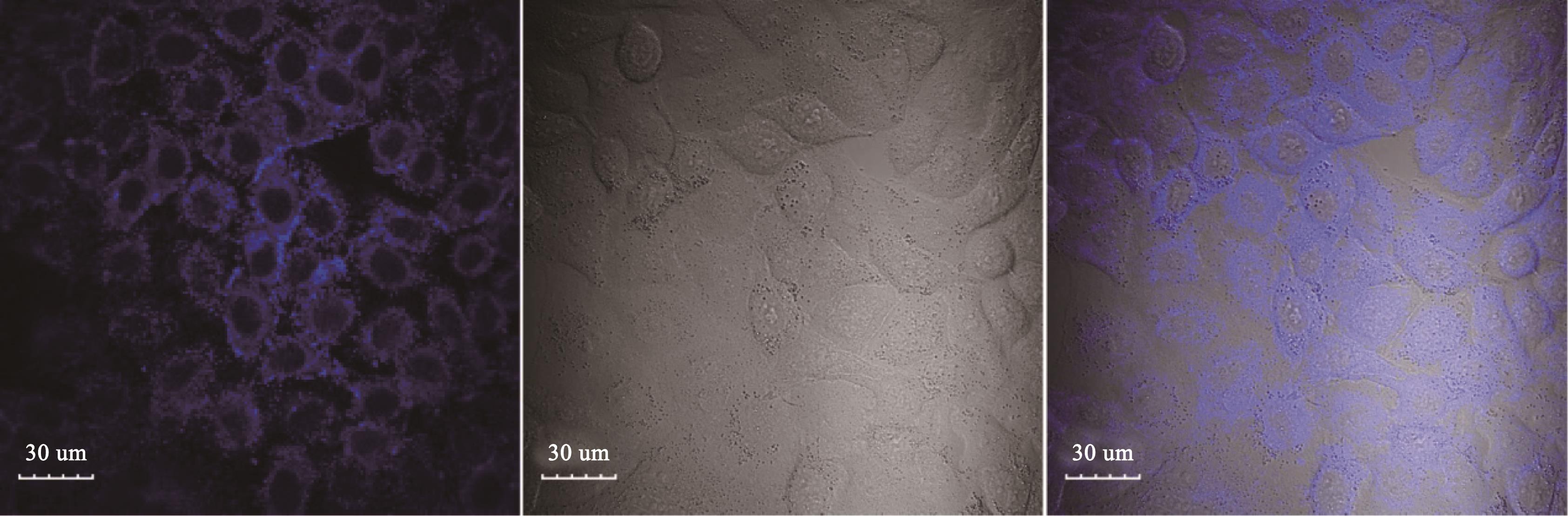
以柠檬酸氢二铵、缓血酸铵和硝酸钕为起始原料,通过水热法合成了荧光量子产率为93.05%、发蓝色荧光的钕、氮双掺杂碳点(Nd,N-CDs)。采用透射电子显微镜(TEM)、粉末X射线衍射(XRD)、红外光谱(FT-IR)及X射线光电子能谱(XPS)等技术对Nd,N-CDs的形貌及表面结构进行了详细表征,采用紫外可见吸收光谱及荧光光谱考察了其光学性质和稳定性。结果显示,药物分子柳氮磺吡啶(SSZ)能够使Nd,N-CDs的荧光显著降低,而金属离子及另外4种药物分子没有明显效果。由此,建立了一种选择性检测SSZ的荧光检测方法,线性检测范围在0.1~50 μmol/L之间,检出限低至0.05 μmol/L。机理探讨表明,荧光猝灭主要涉及动态猝灭和荧光共振能量转移两种机制。所建立的分析方法能用于测定实际药片中的SSZ含量,回收率为98.1%~104.2%。此外,Nd,N-CDs表现出很好的生物相容性,能够作为荧光探针穿透细胞壁使细胞质染色,因不会进一步穿透进入细胞核,很好的避免了对细胞的深层次破坏。

关键词: 碳点, 柳氮磺吡啶, 荧光分析, 细胞成像

Abstract:

Ammonium citrate dibasic, trometamol and neodymium nitrate are employed as raw materials to effectively synthesize blue fluorescent neodymium and nitrogen co-doped carbon dots (Nd,N-CDs) with a fluorescence quantum yield of up to 93.05% via hydrothermal method. The morphology and surface structure of Nd,N-CDs are characterized in detail by several techniques including transmission electron microscopy (TEM), powder X-ray diffraction (XRD), infrared spectroscopy (FT-IR) and X-ray photoelectron spectroscopy (XPS). In addition, ultraviolet visible absorption spectroscopy and fluorescence spectroscopy are used to investigate its optical properties and optical stability. Results show that drug molecule sulfasalazine (SSZ) could dramatic decrease the fluorescence of Nd,N-CDs, but other four drug molecules and metal ions have no significant influence. Thus, a fluorescence detection method for selective detection of SSZ is established. The linear detection range is between 0.1 μmol/L and 50 μmol/L, and the detection limit is 0.05 μmol/L. Further investigation shows that the fluorescence quenching mainly involves two mechanisms that are dynamic quenching and fluorescence resonance energy transfer. The as-established analytical method could be used to determine the content of SSZ in actual tablets with recovery ranging from 98.1%~104.2%. Furthermore, the synthesized Nd,N-CDs present good biocompatibility and could be used as a fluorescent probe to penetrate the cell wall to stain the cytoplasm, however, it could not enter the cell nucleus, which effectively avoids further cell damage.

Key words: Carbon dots, Sulfasalazine, Fluorescence analysis, Cell imaging

中图分类号: