Chinese Journal of Applied Chemistry ›› 2025, Vol. 42 ›› Issue (8): 1035-1056.DOI: 10.19894/j.issn.1000-0518.250087
• Review • Next Articles
Xin-Xin HUANG1, You-Sheng SHI2, Tao DENG3, Chun CAI1( )
)
Received:2025-03-01
Accepted:2025-06-11
Published:2025-08-01
Online:2025-08-11
Contact:
Chun CAI
About author:c.cai@njust.edu.cnCLC Number:
Xin-Xin HUANG, You-Sheng SHI, Tao DENG, Chun CAI. Research Progress of Fluorescent Probes Based on Aggregation-Induced Emission for Detection of Biomarkers[J]. Chinese Journal of Applied Chemistry, 2025, 42(8): 1035-1056.
Add to citation manager EndNote|Ris|BibTeX
URL: http://yyhx.ciac.jl.cn/EN/10.19894/j.issn.1000-0518.250087
| Diseases | Biomarkers |
|---|---|
| Lung cancer | Carcinoembryonic antigen (CEA), neuron specific enolase (NSE), squamous cell carcinoma antigen (SCCA), cytokeratin 19 fragment antigen (CYFRA21-1), biothiols etc. |
| Liver cancer | Alpha fetoprotein (AFP), alpha-L fucosidase (AFU), hepatocyte growth factor (HGF) etc. |
| Gastric cancer | Carcinoembryonic antigen (CEA), carbohydrate antigen (CA 19-9, CA72-4), pepsinogen Ⅰ/Ⅱ(PG Ⅰ/Ⅱ) etc. |
| Breast cancer | Carcinoembryonic antigen (CEA), carbohydrate antigen (CA), tissue polypeptide specific antigen (TPS), epithelial cadherin (E-cadherin), alkaline phosphatase (ALP), nitroreductase (NTR), H2S etc. |
| Cervical cancer | Carbohydrate antigen (CA125,CA72-4), squamous cell carcinoma antigen (SCCA), hepatocyte growth factor (HGF), mixed lineage kinase domain-like protein (MLKL), Cu2+, Hg2+etc. |
| Ovary cancer | Tumor necrosis factor-α (TNF-α), humanepididymisprotein4 (HE4), hexokinase2 (HK2), sequestosome-1 (SQSTM1/p62),β-galactosidase (β-Gal) etc. |
| Colorectal cancer | Carcinoembryonic antigen (CEA), lactate dehydrogenase (LDH), microtubule-associated protein 1 light chain 3 beta (MAP1LC3B), unc-51 like autophagy activating kinase 1 (ULK-1), glutathione (GSH) etc. |
| Cardiovascular disease | Myoglobin (Myo), cardiac troponin (cTn), creatine kinase isoenzyme (CK-MB), B-type natriuretic peptide (BNP), growth differentiation factor 15 (GDF-15) etc. |
| Alzheimer's disease | Amyloid-β peptides (Aβ peptides), tau protein, apolipoprotein E (apoE), circulating free microRNAs (miRNAs), alpha-1 antitrypsin (AAT), Zn2+etc. |
| Inflammation | C-reactive protein (CRP), serum amyloid A (SAA), procalcitonin (PCT), ferritin (SF), reactive oxygen species (ROS) etc. |
Table 1 Examples of universally acknowledged biomarkers and related diseases
| Diseases | Biomarkers |
|---|---|
| Lung cancer | Carcinoembryonic antigen (CEA), neuron specific enolase (NSE), squamous cell carcinoma antigen (SCCA), cytokeratin 19 fragment antigen (CYFRA21-1), biothiols etc. |
| Liver cancer | Alpha fetoprotein (AFP), alpha-L fucosidase (AFU), hepatocyte growth factor (HGF) etc. |
| Gastric cancer | Carcinoembryonic antigen (CEA), carbohydrate antigen (CA 19-9, CA72-4), pepsinogen Ⅰ/Ⅱ(PG Ⅰ/Ⅱ) etc. |
| Breast cancer | Carcinoembryonic antigen (CEA), carbohydrate antigen (CA), tissue polypeptide specific antigen (TPS), epithelial cadherin (E-cadherin), alkaline phosphatase (ALP), nitroreductase (NTR), H2S etc. |
| Cervical cancer | Carbohydrate antigen (CA125,CA72-4), squamous cell carcinoma antigen (SCCA), hepatocyte growth factor (HGF), mixed lineage kinase domain-like protein (MLKL), Cu2+, Hg2+etc. |
| Ovary cancer | Tumor necrosis factor-α (TNF-α), humanepididymisprotein4 (HE4), hexokinase2 (HK2), sequestosome-1 (SQSTM1/p62),β-galactosidase (β-Gal) etc. |
| Colorectal cancer | Carcinoembryonic antigen (CEA), lactate dehydrogenase (LDH), microtubule-associated protein 1 light chain 3 beta (MAP1LC3B), unc-51 like autophagy activating kinase 1 (ULK-1), glutathione (GSH) etc. |
| Cardiovascular disease | Myoglobin (Myo), cardiac troponin (cTn), creatine kinase isoenzyme (CK-MB), B-type natriuretic peptide (BNP), growth differentiation factor 15 (GDF-15) etc. |
| Alzheimer's disease | Amyloid-β peptides (Aβ peptides), tau protein, apolipoprotein E (apoE), circulating free microRNAs (miRNAs), alpha-1 antitrypsin (AAT), Zn2+etc. |
| Inflammation | C-reactive protein (CRP), serum amyloid A (SAA), procalcitonin (PCT), ferritin (SF), reactive oxygen species (ROS) etc. |
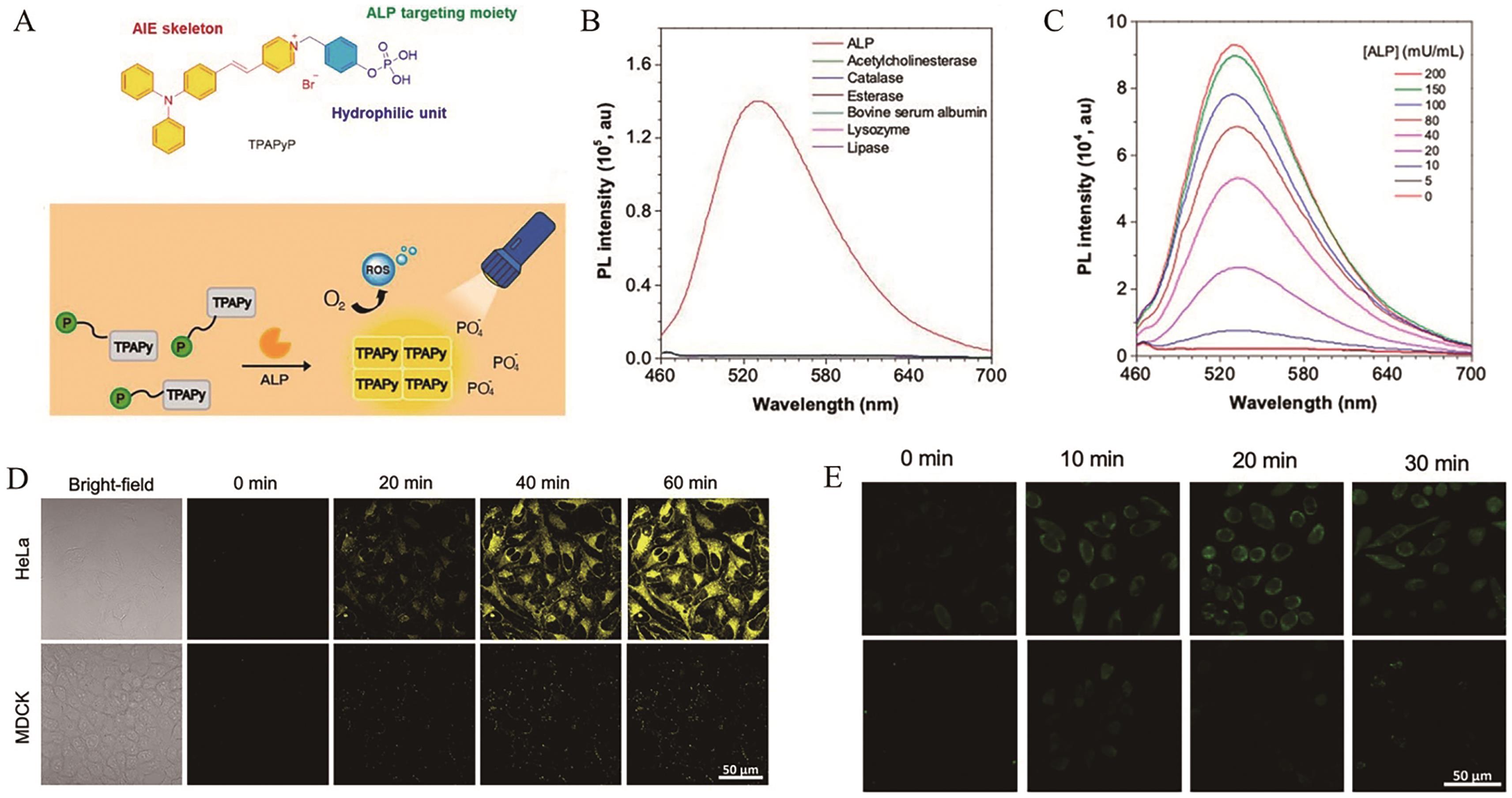
Fig.1 (A) The structure and response mechanism of AIE probe TPAPyP; (B) The fluorescence response of probe TPAPyP (100 μmol/L) after incubation with different analytes; (C) The fluorescence spectra of probe TPAPyP (100 μmol/L) after reaction with various concentrations of ALP; (D) The fluorescence imaging of HeLa cells and MDCK cells incubated with probe TPAPyP (2 μmol/L); (E) The fluorescence imaging of HeLa cells (top) and MDCK cells (bottom) incubated with ROS probe DCFH[34]
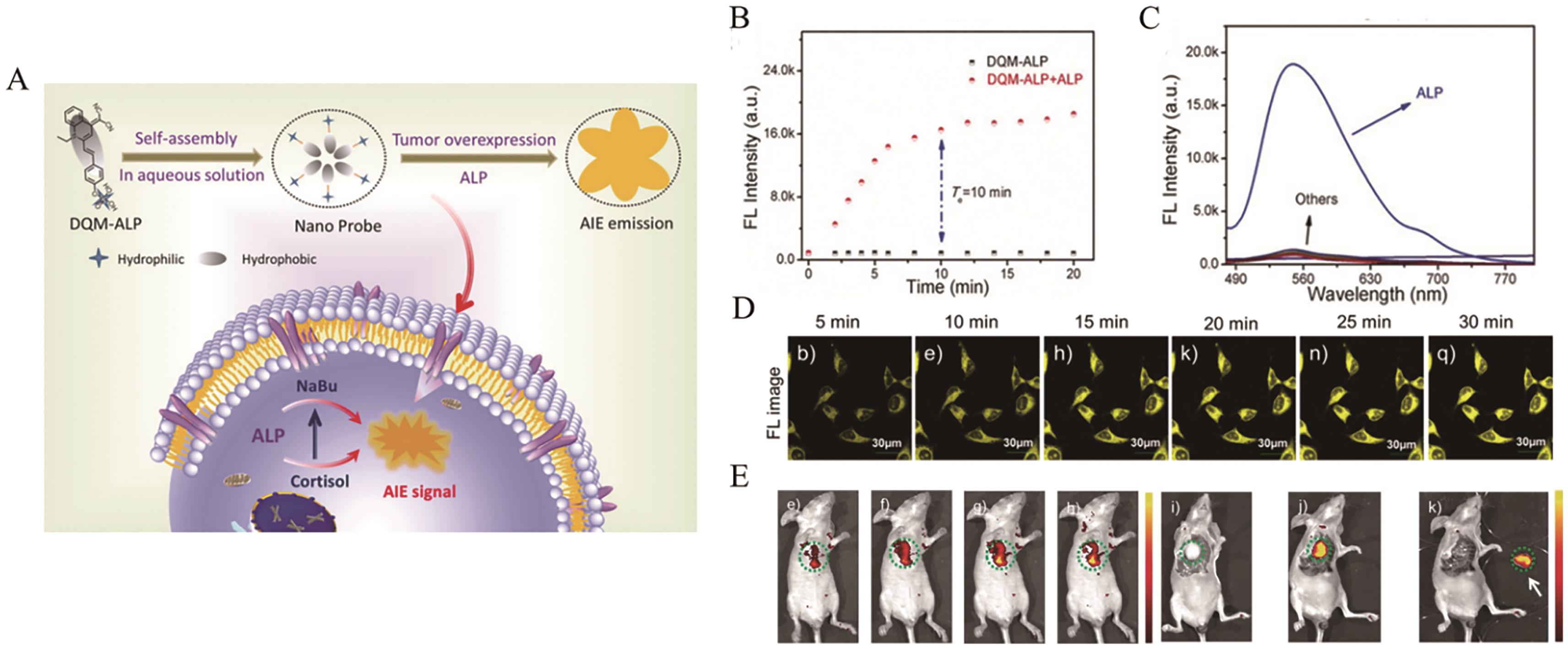
Fig.2 (A) The structure and schematic illustration of intracellular response of AIE probe DQM-ALP; (B) The kinetic curves of probe DQM-ALP before and after response to ALP; (C) The fluorescence response of probe DQM-ALP (10 μmol/L) after incubation with different analytes; (D) The fluorescence imaging of HeLa cells incubated with probe DQM-ALP (10 μmol/L); (E) Fluorescence imaging of liver tumor-bearing mice after injection of probe DQM-ALP (50 μmol/L)[35]
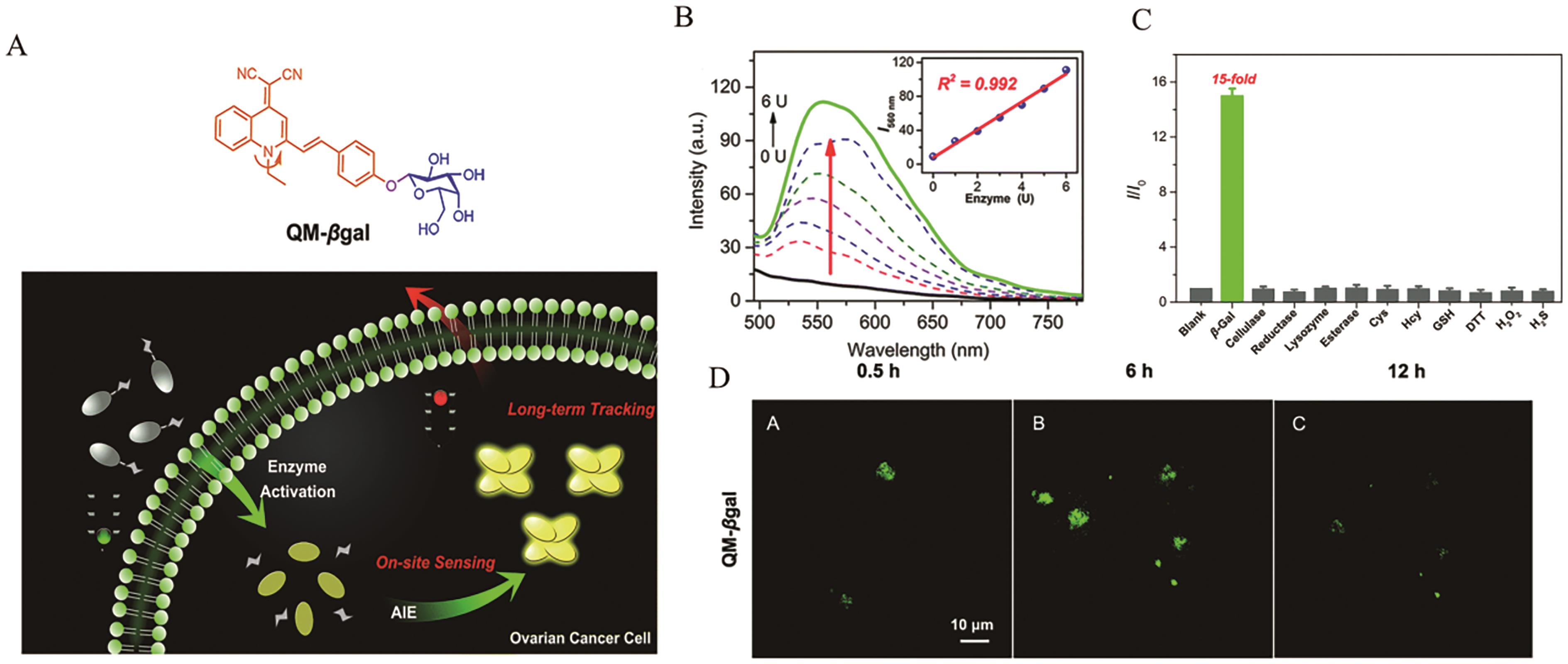
Fig.3 (A) The structure and schematic illustration of intracellular response of AIE probe QM-βgal; (B) The fluorescence spectra of probe QM-βgal (10 μmol/L) after reaction with various concentrations of β-Gal and corresponding linear relationship; (C) The fluorescence response of probe QM-βgal (10 μmol/L) after incubation with different analytes; (D) The fluorescence imaging of SKOV-3 cells incubated with probe QM-βgal (10 μmol/L)[37]
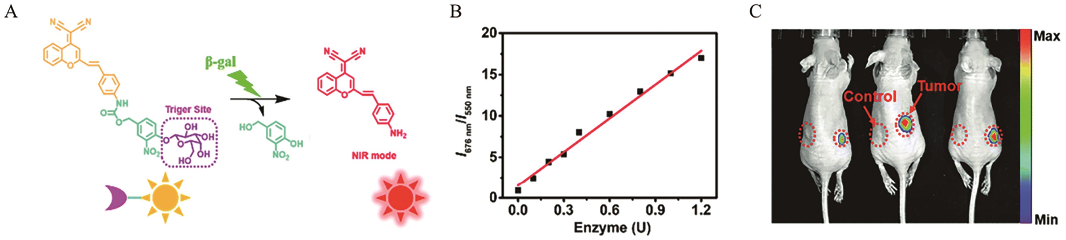
Fig.4 (A) The structure and response mechanism of AIE probe DCMCA-β-gal; (B) The linear relationship between the ratio-metric fluorescence intensity I676 nm/I550 nm of probe DCMCA-β-gal and the concentration of β-Gal; (C) Fluorescence imaging of ovarian tumor-bearing mice after injection of probe DCMCA-β-gal[38]
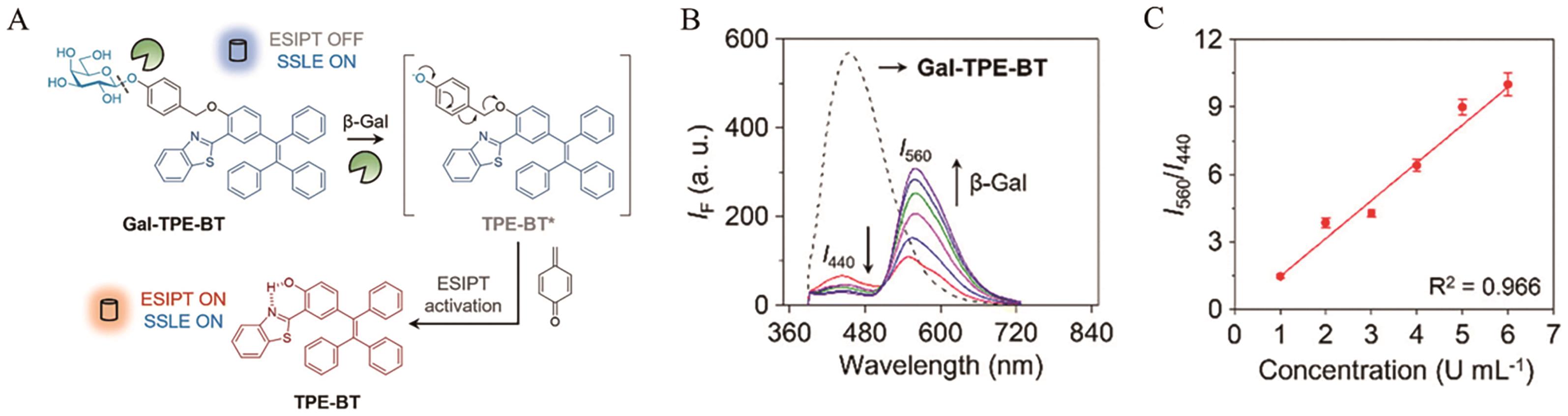
Fig.5 (A)The structure and response mechanism of AIE probe Gal-TPE-BT; (B) The fluorescence spectra of probe Gal-TPE-BT (10 μmol/L) after reaction with various concentrations of β-Gal; (C) The linear relationship between the ratio-metric fluorescence intensity I550 nm/I440 nm of probe Gal-TPE-BT and the concentration of β-Gal[39]
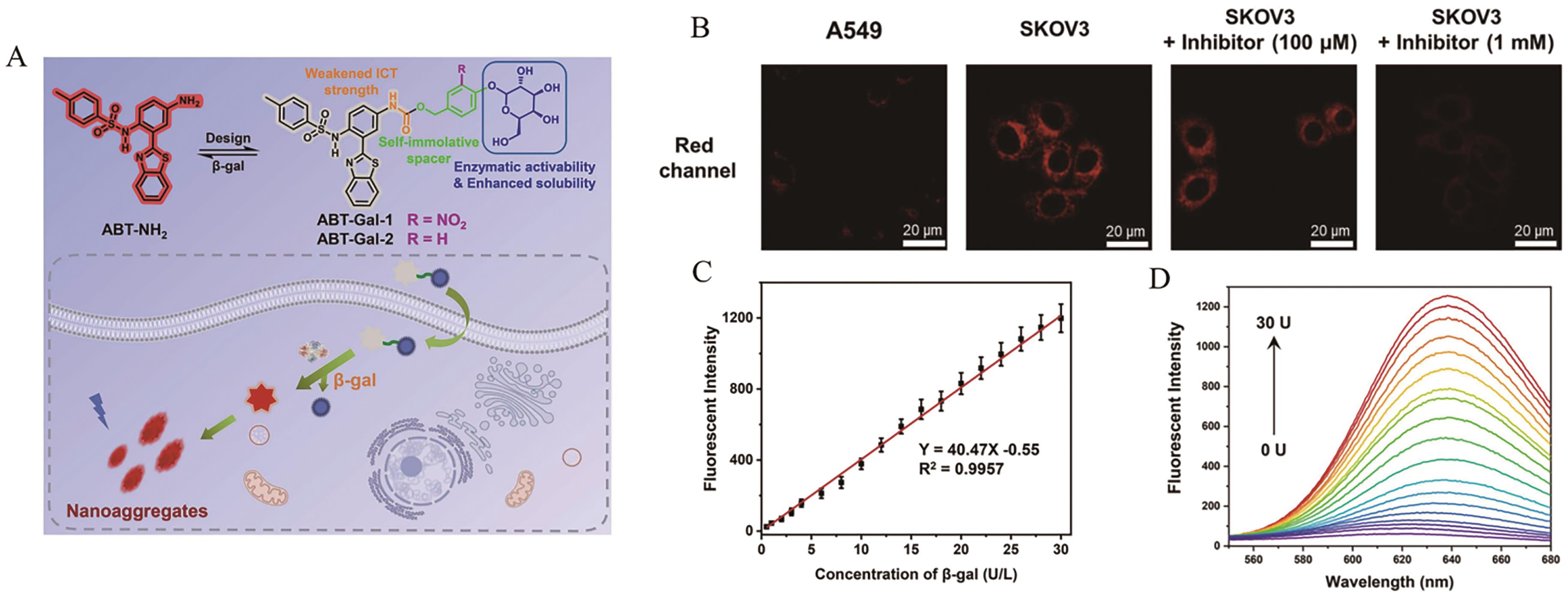
Fig.6 (A) The structure and schematic illustration of intracellular response of AIE probe ABT-Gal; (B) The fluorescence imaging of A549 cells and SKOV-3 cells incubated with probe ABT-Gal-1 (20 μmol/L); (C) The linear relationship between the fluorescence intensity of probe ABT-Gal-1 and the concentration of β-Gal; (D) The fluorescence spectra of probe ABT-Gal-1 (10 μmol/L) after reaction with various concentrations of β-Gal[40]
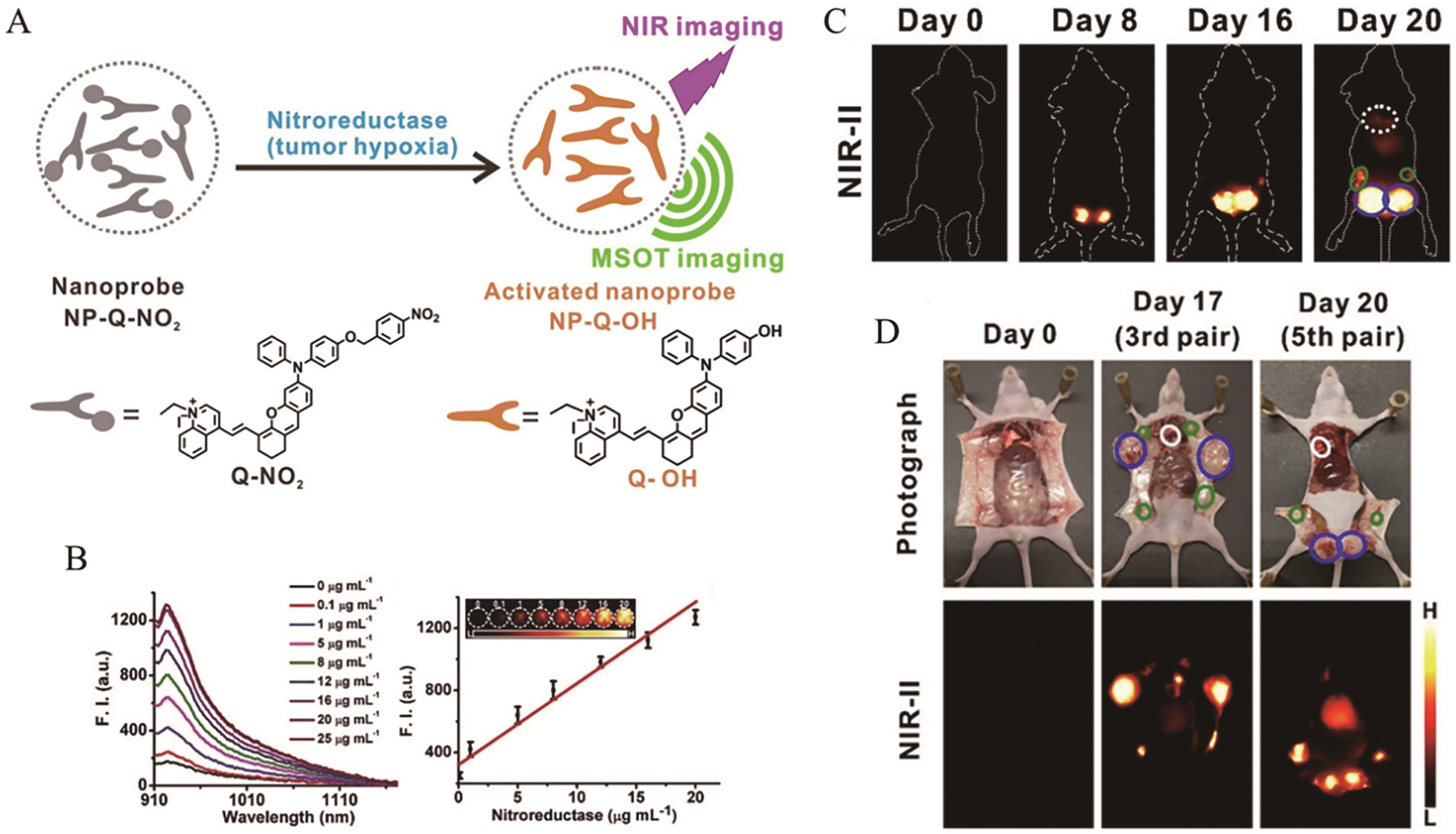
Fig.7 (A) The structure and response mechanism of AIE probe Q-NO2; (B) The fluorescence spectra of probe Q-NO2 (10 μmol/L) after reaction with various concentrations of NTR and corresponding linear relationship; (C) Fluorescence imaging of breast tumor-bearing mice after injection of probe Q-NO2; (D) Fluorescence imaging of tumor-bearing mice after injection of probe Q-NO2[41]
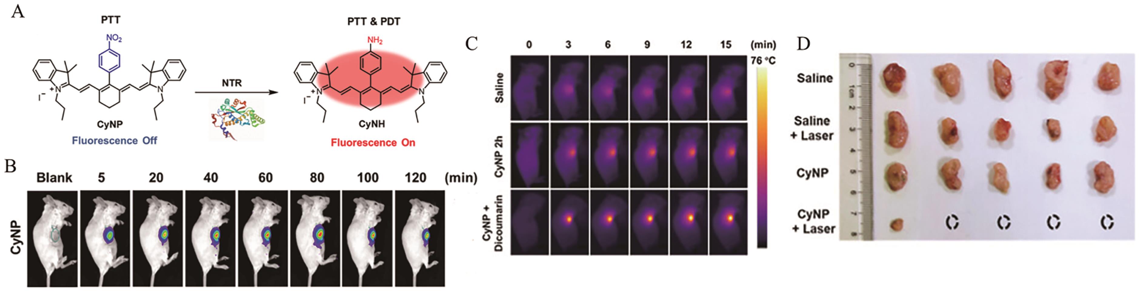
Fig.8 (A) The structure and response mechanism of AIE probe CyNP; (B) Fluorescence imaging of breast tumor-bearing mice after injection of probe CyNP; (C) Photothermal imaging of breast tumor-bearing mice after injection of probe CyNP; (D) Photos of tumor in mice[42]
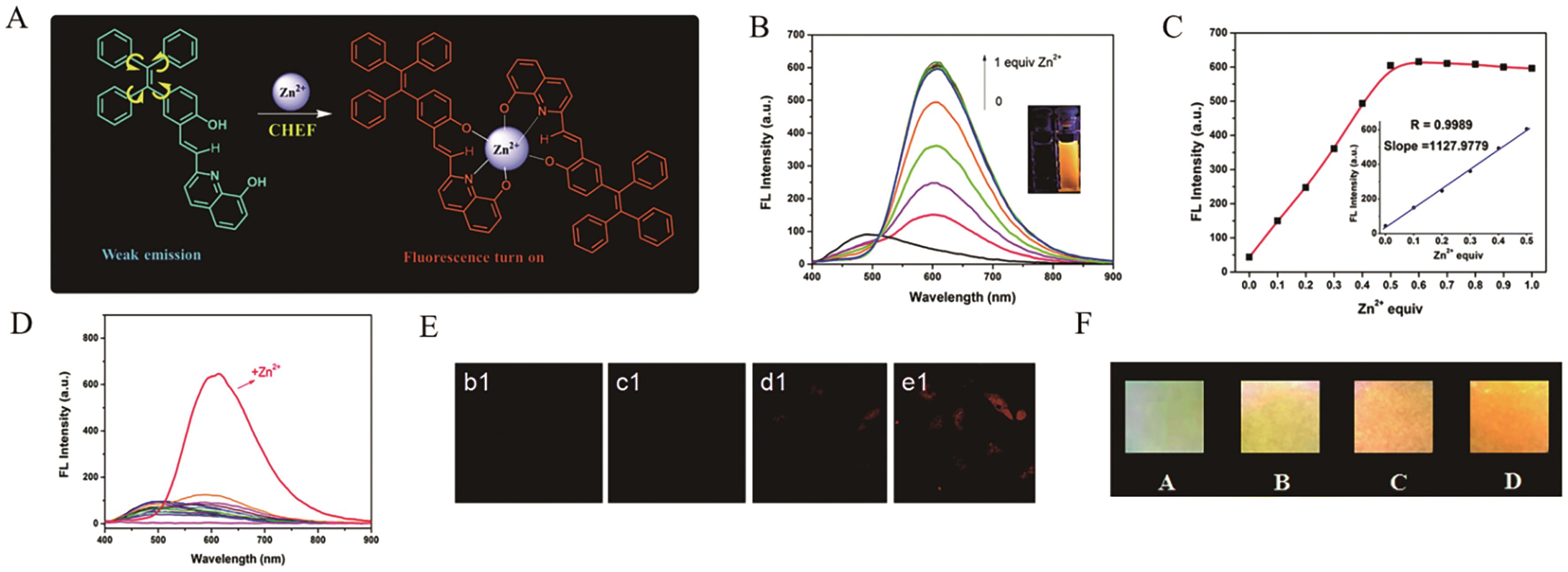
Fig.9 (A) The structure and response mechanism of AIE probe TPE(OH)-8HQ; (B) The fluorescence spectra of probe TPE(OH)-8HQ (10 μmol/L) after reaction with various concentrations of Zn2+; (C) The linear relationship between the fluorescence intensity of probe TPE(OH)-8HQ and the concentration of Zn2+; (D) The fluorescence response of probe TPE(OH)-8HQ (10 μmol/L) after incubation with different analytes; (E) The fluorescence imaging of HeLa cells incubated with probe TPE(OH)-8HQ (20 μmol/L); (F) Fluorescence response of TPE(OH)-8HQ (1 mmol/L) to Zn2+ on test papers[47]
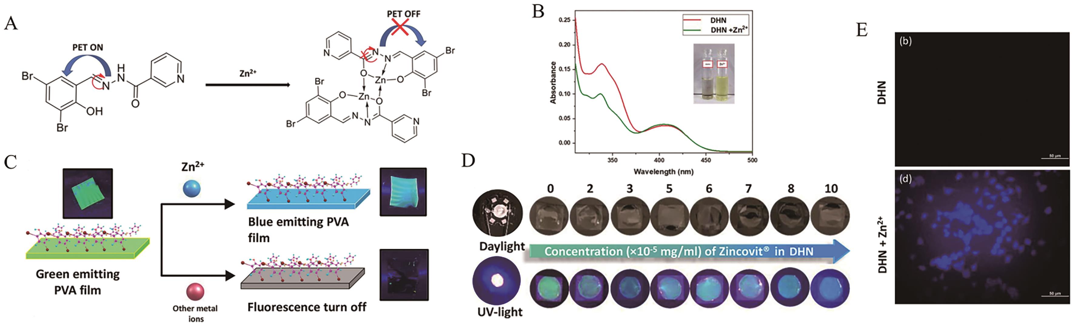
Fig.10 (A) The structure and response mechanism of AIE probe DHN; (B) The absorption spectra of probe DHN(10 μmol/L) before and after response to Zn2+; (C) The detection mechanism of PVA_DHN composite film; (D) Photos of DHN response to various concentrations of Zincovit under UV irradiation; (E) The fluorescence imaging of N2a cells incubated with probe DHN (5 μmol/L)[48]
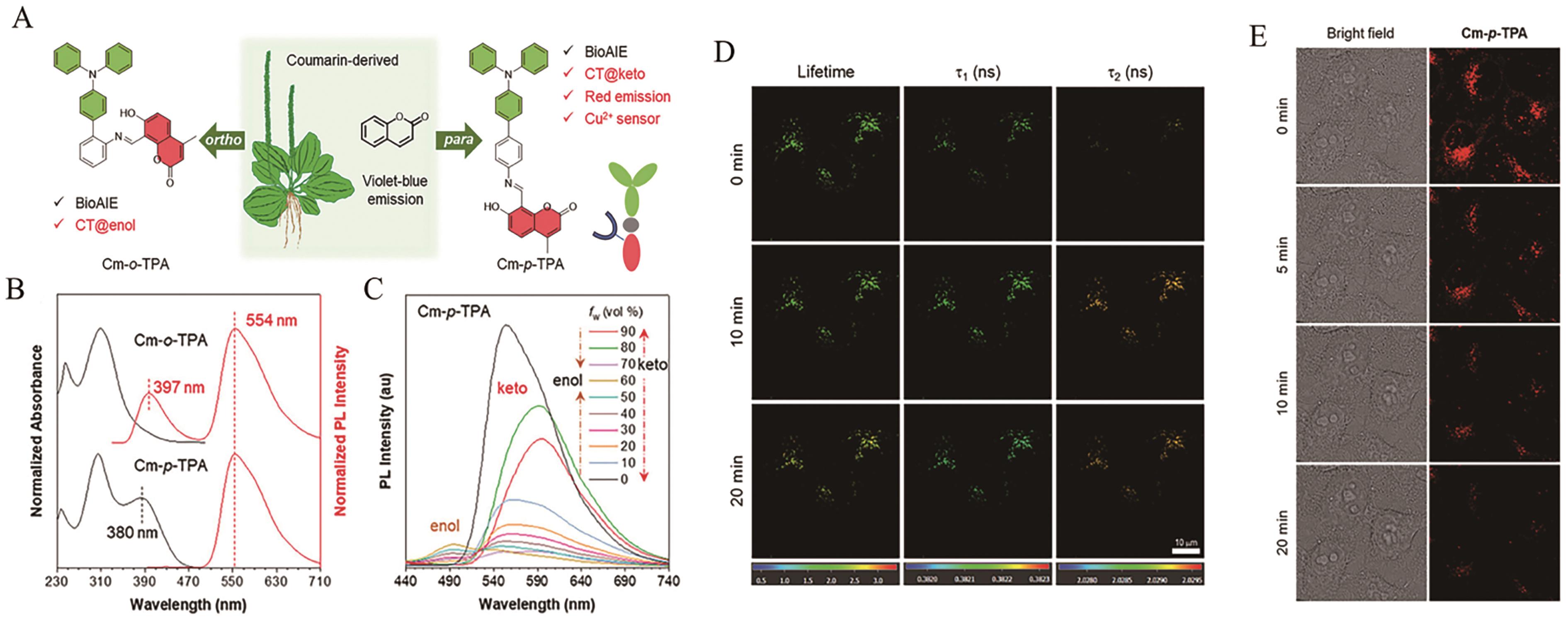
Fig.11 (A) The design strategy of AIE probe Cm-o-TPA and Cm-p-TPA; (B) The normalized absorption spectra of Cm-o-TPA and Cm-p-TPA in tetrahydrofuran (THF) solution; (C) The fluorescence spectra of Cm-p-TPA in THF/H2O mixed solvent with different water fractions (fw); (D) The lifetime changes of Cm-p-TPA NPs (10 μmol/L) in HeLa cells; (E) Monitoring of mitophagy process with Cm-p-TPA NPs (10 μmol/L)[51]
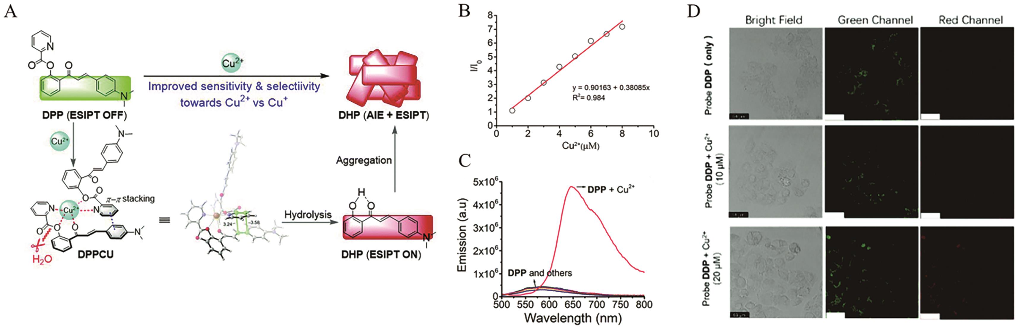
Fig.12 (A) The structure and response mechanism of AIE probe DPP; (B) The linear relationship between the relative fluorescence intensity I648 nm/I0 of probe DPP and the concentration of Cu2+; (C) The fluorescence response of probe DPP (20 μmol/L) after incubation with different analytes; (D) The fluorescence imaging of HeLa cells incubated with probe DPP(10 μmol/L)[52]
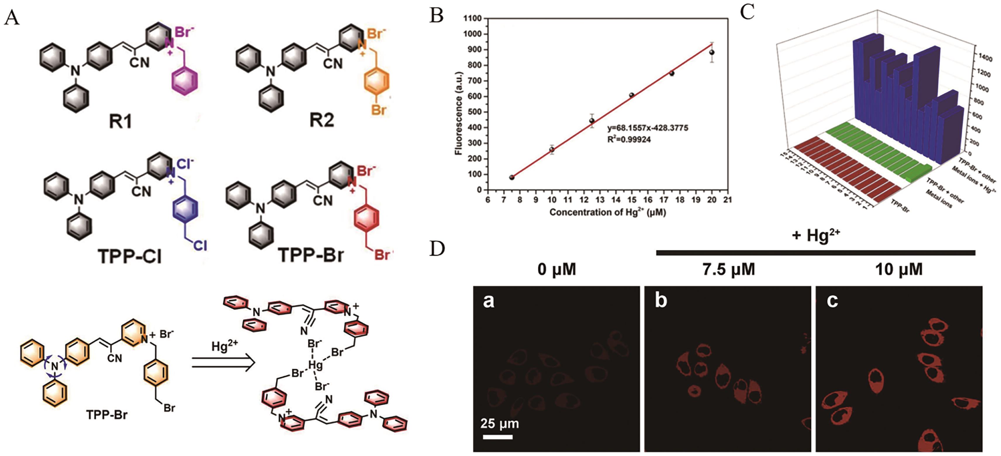
Fig.13 (A) The structure of AIE probe R1, R2, TPP-Cl and TPP-Br and response mechanism of TPP-Br; (B) The linear relationship between the fluorescence intensity of probe TPP-Br and the concentration of Hg2+; (C) The fluorescence response of probe TPP-Br (50 μmol/L) after incubation with different analytes in the presence of Hg2+ (30?μmol/L); (D) The fluorescence imaging of HeLa cells incubated with probe TPP-Br (10 μmol/L)[57]
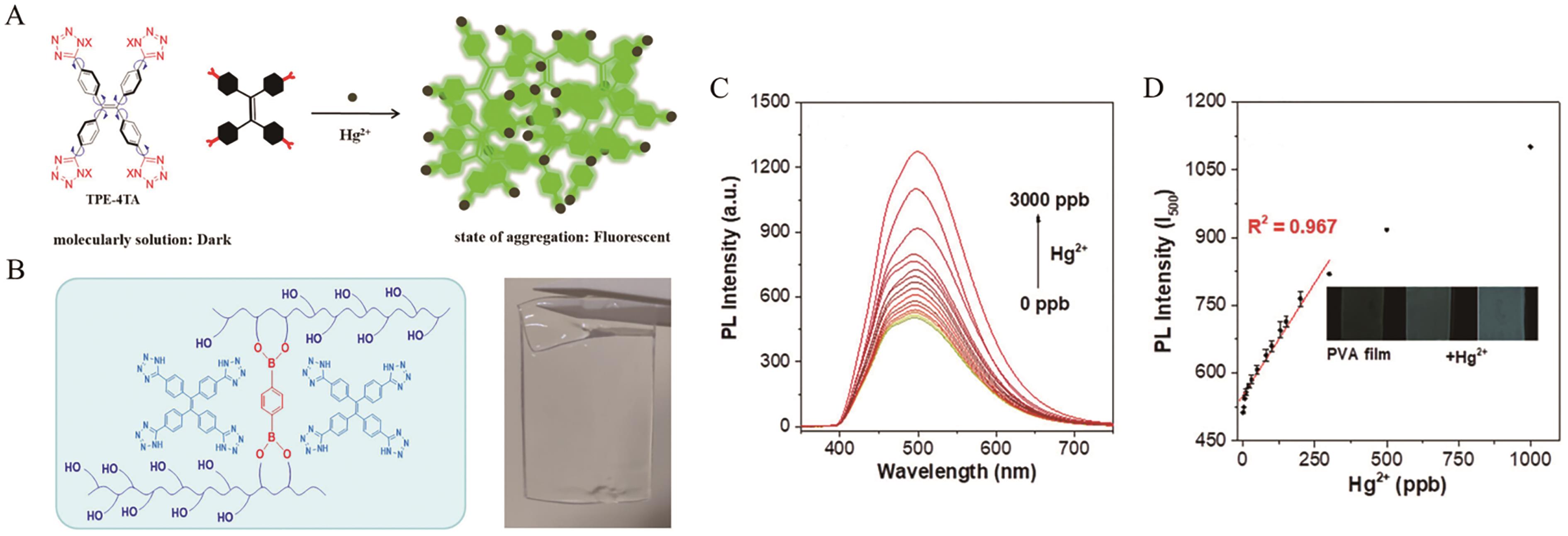
Fig.14 (A) The structure and response mechanism of AIE probe TPE-4TA; (B) The polyvinyl alcohol (PVA)-based hydrogel film dopped with TPE-4TA; (C) The fluorescence spectra of PVA-based hydrogel film after reaction with various concentrations of Hg2+; (D) The linear relationship between the fluorescence intensity of PVA-based hydrogel film and the concentration of Hg2+[58]
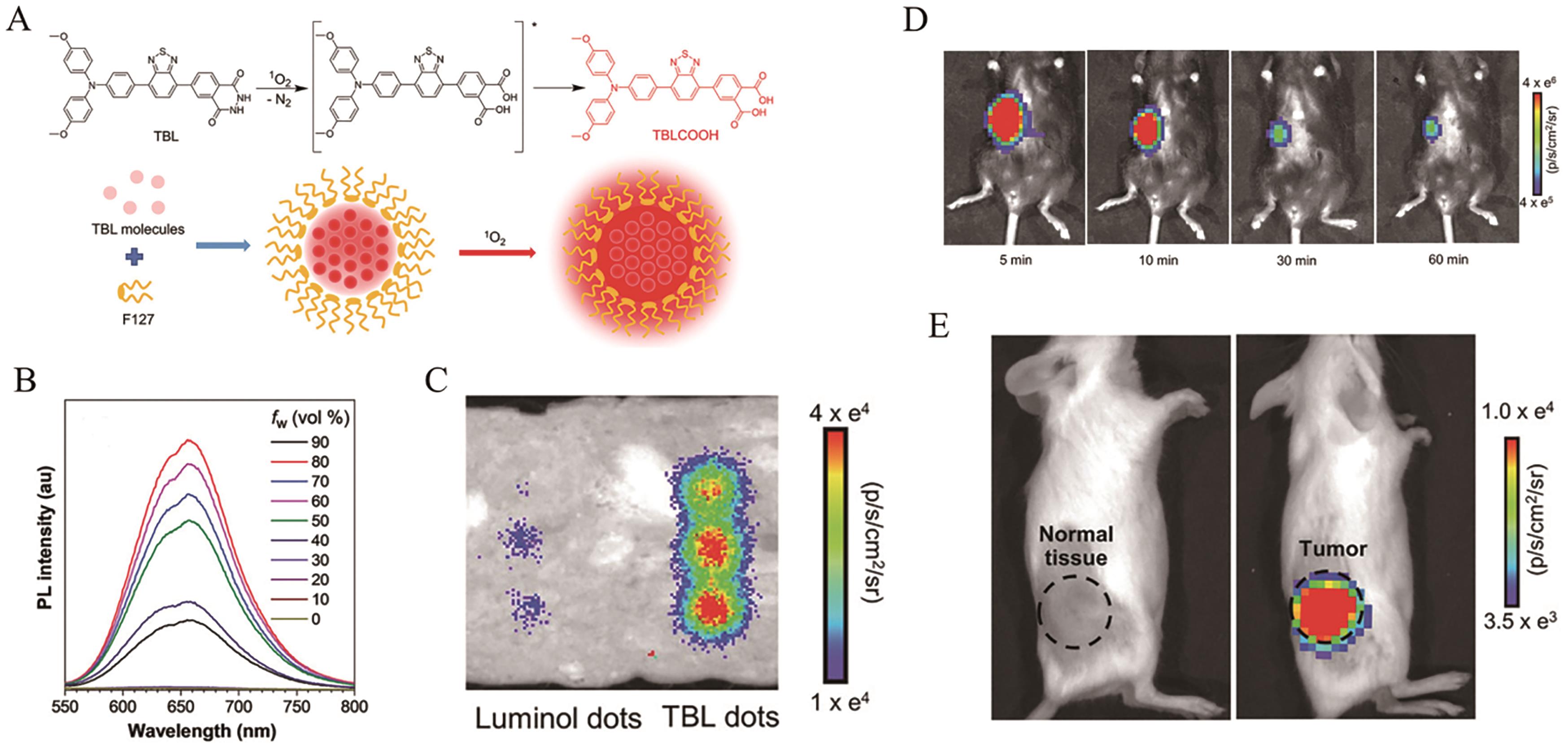
Fig.15 (A) The structure and response mechanism of AIE probe TBL; (B) The fluorescence spectra of TBL in dimethyl sulfoxide (DMSO)/H2O mixed solvent with different water fractions (fw); (C) Chemiluminescence of luminol dots (1.2 mmol/L) and TBL dots (1.2 mmol/L) covered by a piece of approximately 3 mm thick pork ham in a H2O2/NaClO mixed solution; (D) The chemiluminescence imaging of mice after subcutaneous injection of TBL dots with H2O2 and NaClO; (E) The chemiluminescence imaging of breast tumor-bearing mice after injection of TBL dots[65]
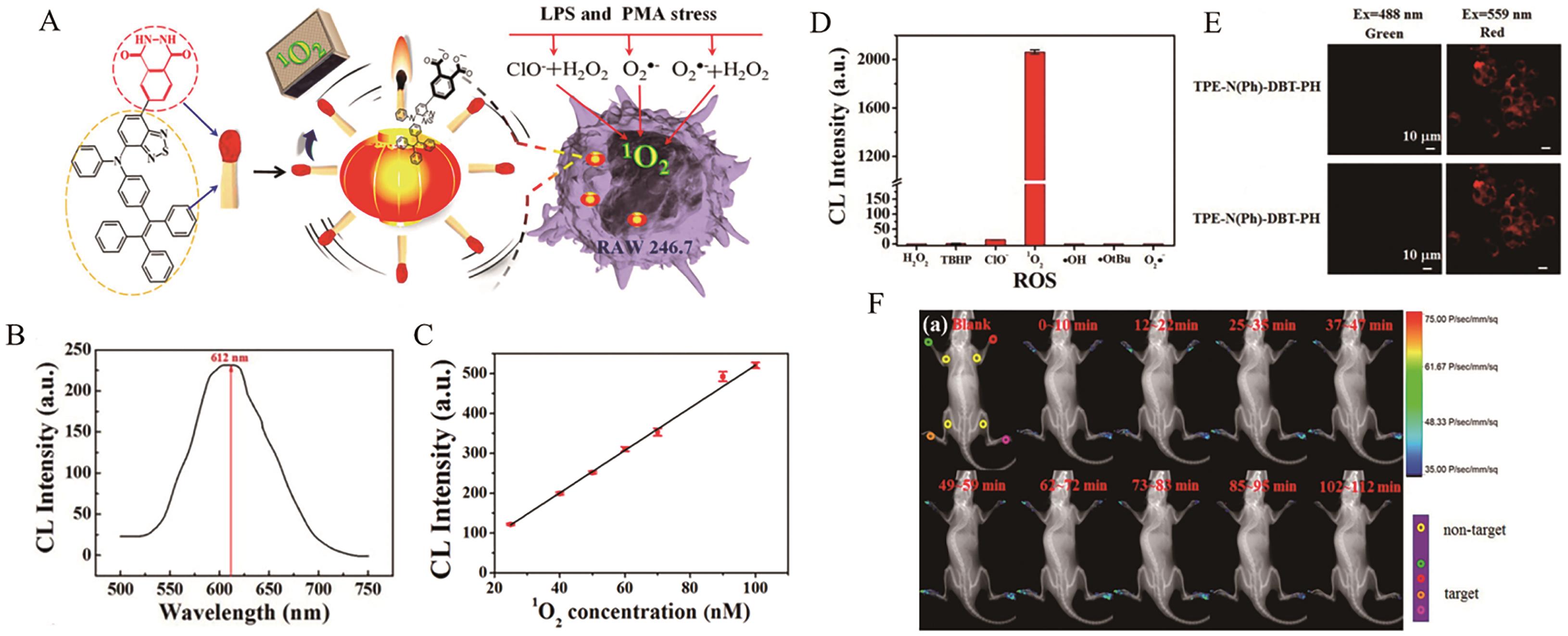
Fig.16 (A) The structure and response mechanism of AIE probe TPE-N(Ph)-DBT-PH; (B) The chemiluminescence spectra of TPE-N(Ph)-DBT-PH; (C) The linear relationship between the chemiluminescence intensity of probe TPE-N(Ph)-DBT-PH and the concentration of 1O2; (D) The chemiluminescence response of probe TPE-N(Ph)-DBT-PH (10 μmol/L) after incubation with different analytes; (E) The chemiluminescence imaging of RAW 264.7 cells incubated with probe TPE-N(Ph)-DBT-PH; (F) The chemiluminescence imaging of a arthritismouse model after injection of TPE-N(Ph)-DBT-PH[66]
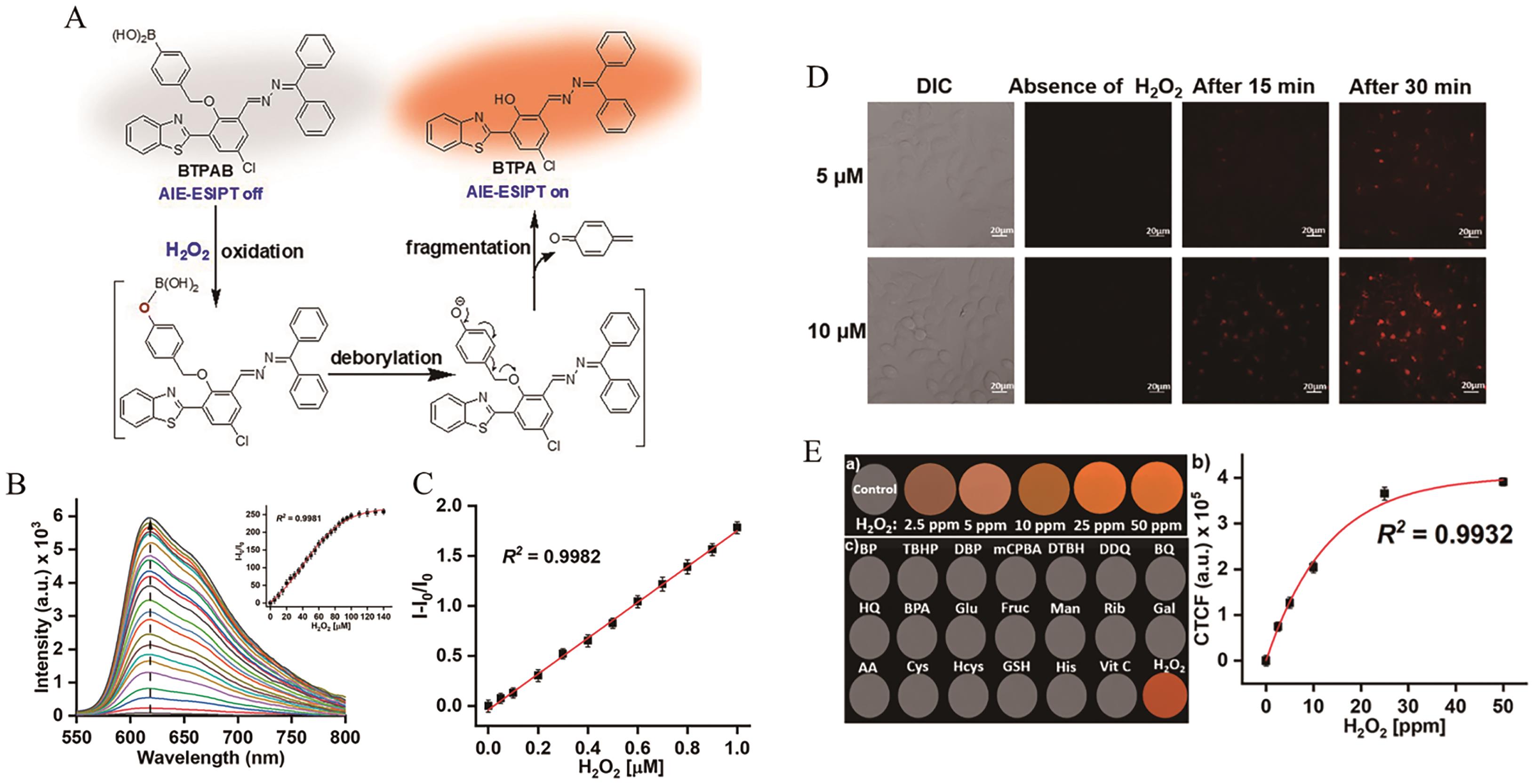
Fig.17 (A) The structure and response mechanism of AIE probe BTPAB; (B) The fluorescence spectra of BTPAB(10 μmol/L) after reaction with various concentrations of H2O2; (C) The linear relationship between the fluorescence intensity of BTPAB and the concentration of H2O2; (D) The fluorescence imaging of HeLa cells incubated with probe BTPAB; (E) Fluorescence response of BTPAB to H2O2 on TLC plates and corresponding linear relationship[69]
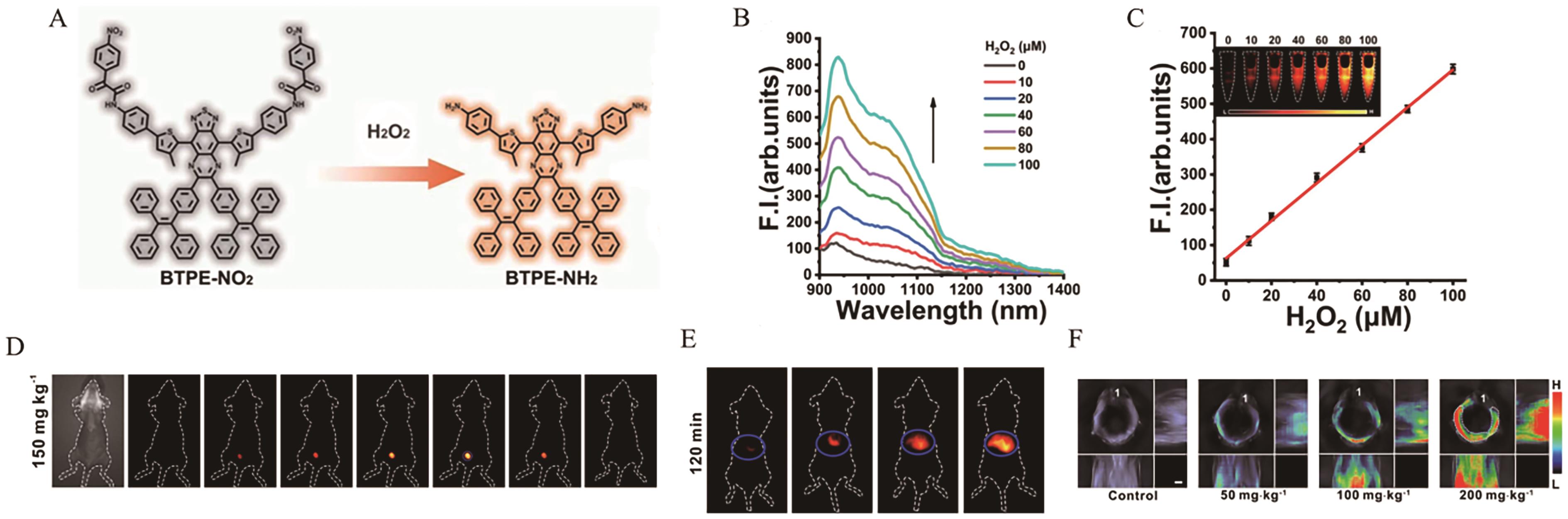
Fig.18 (A) The structure and response mechanism of AIE probe BTPE-NO2; (B) The fluorescence spectra of BTPE-NO2@F127 (BTPE-NO2 32.6?μg/mL) after reaction with various concentrations of H2O2; (C) The linear relationship between the fluorescence intensity of BTPE-NO2@F127 and the concentration of H2O2; (D) The fluorescence imaging of an interstitial cystitis mouse model after injection of BTPE-NO2@F127; (E) The fluorescence imaging of a trazodone-induced liver injury mouse model after injection of BTPE-NO2@F127; (F) The multispectral optoacoustic tomography imaging of a trazodone-induced liver injury mouse model after injection of BTPE-NO2@F127[70]
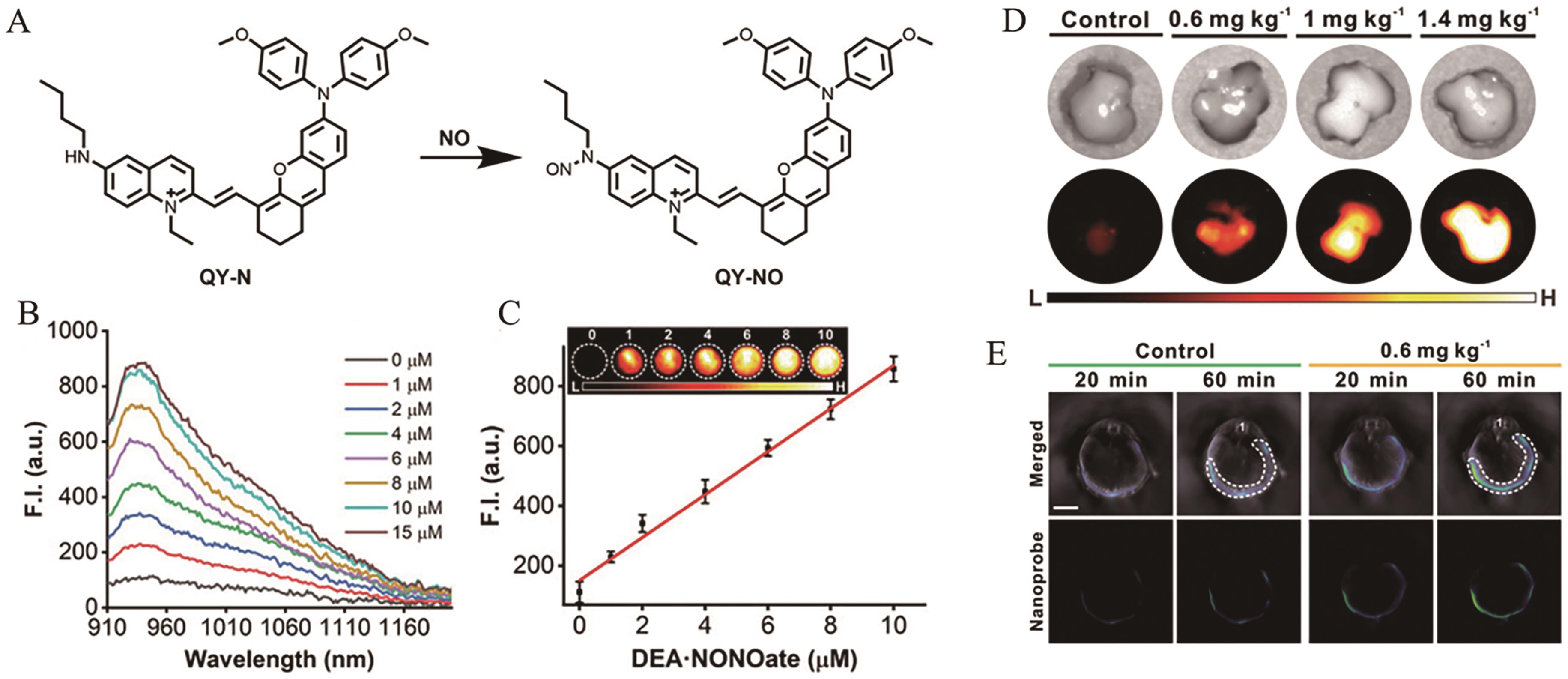
Fig.19 (A) The structure and response mechanism of AIE probe QY-N; (B) The fluorescence spectra of QY-N (10 μmol/L) after reaction with various concentrations of NO; (C) The linear relationship between the fluorescence intensity of QY-N and the concentration of NO; (D) The fluorescence imaging of livers incubated with probe QY-N; (E) The multispectral optoacoustic tomography imaging of livers after injection of QY-N[73]
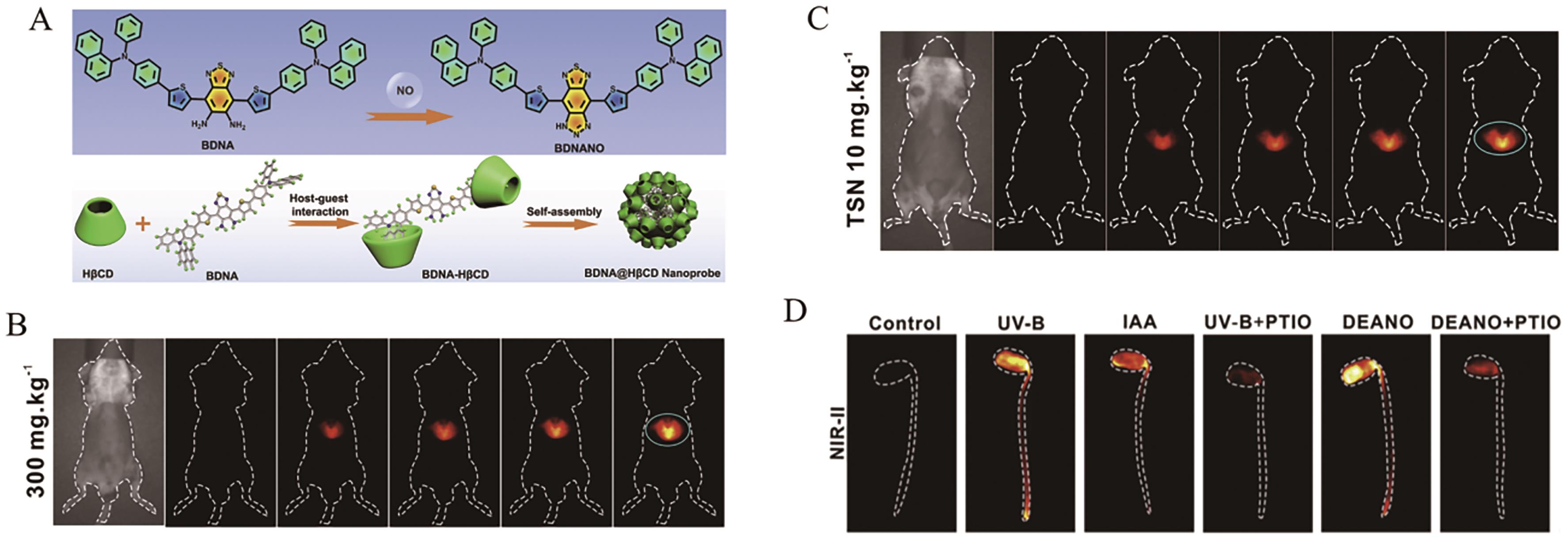
Fig.20 (A) The structure and response mechanism of AIE probe BNDA; (B) The fluorescence imaging of an APAP-induced liver injury mouse model after injection of BNDA@HβCD; (C) The fluorescence imaging of an TSN-induce dliver injury mouse model after injection of BNDA@HβCD; (D) The fluorescence imaging of soybean sprouts[74]

Fig.21 (A) The structure and response mechanism of AIE probe DNBS-HCA; (B) The fluorescence spectra of DNBS-HCA after reaction with various concentrations of GSH, Cys and Hcy, repectively; (C) The linear relationship between the fluorescence intensity of DNBS-HCA and the concentration of GSH, Cys and Hcy, repectively; (D) The fluorescence imaging of PC-3 cells and A549 cells incubated with probe DNBS-HCA; (E) Fluorescence response of DNBS-HCA to Cys on test papers[77]
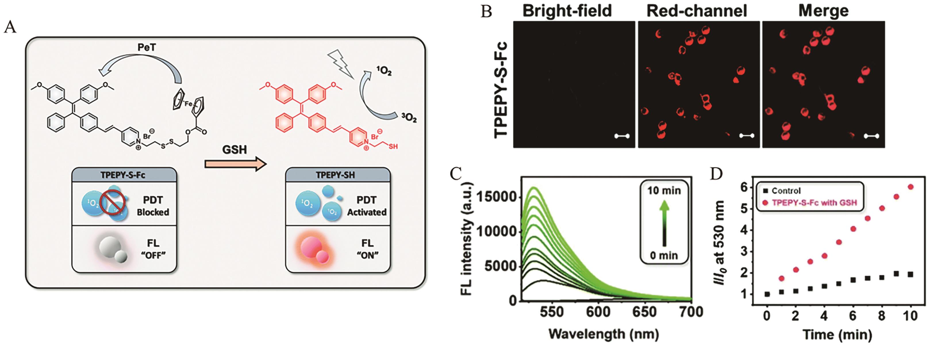
Fig.22 (A) The structure and response mechanism of AIE probe TPEPY-S-Fc; (B) The fluorescence imaging of CT-26 cells incubated with probe TPEPY-S-Fc; (C) The fluorescence spectra of 1O2 probe SOSG in the presence of TPEPY-S-Fc (20 μmol/L) and GSH (200 μmol/L); (D) The time-dependent relative fluorescence intensity I530 nm/I0 of probe SOSG[78]
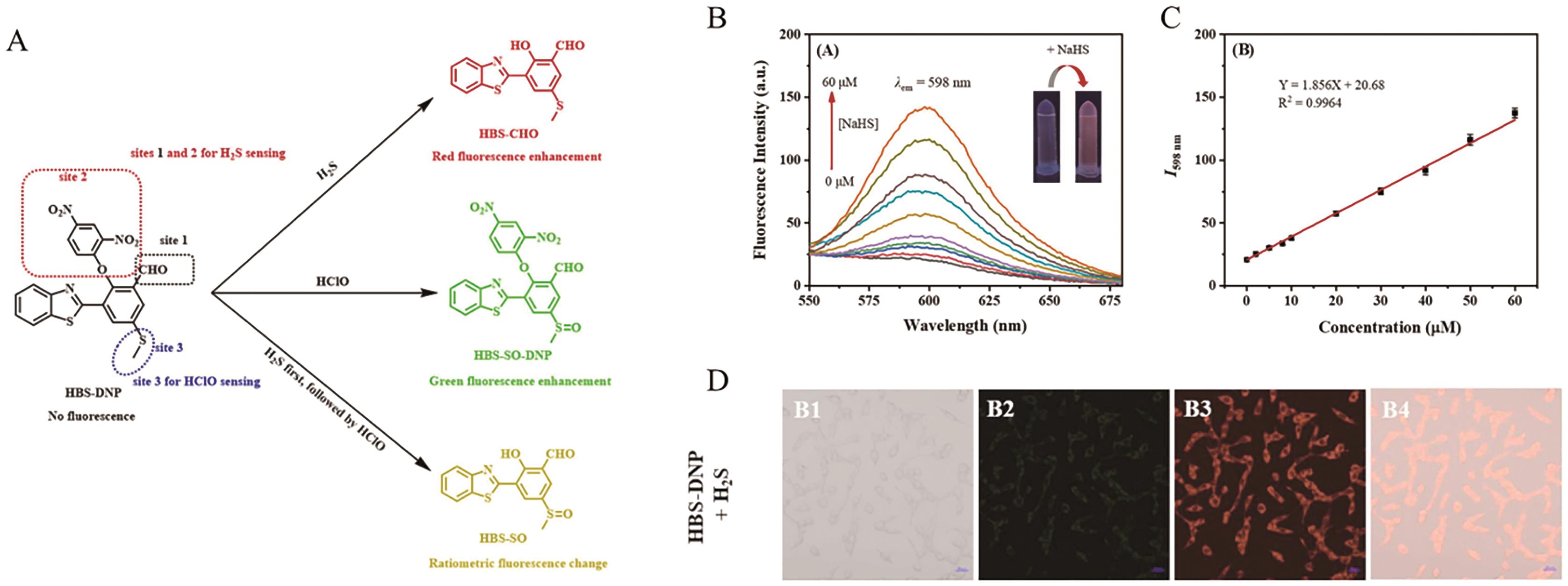
Fig.23 (A) The structure and response mechanism of AIE probe HBS-DNP; (B) The fluorescence spectra of HBS-DNP (10 μmol/L) after reaction with various concentrations of H2S; (C) The linear relationship between the fluorescence intensity of HBS-DNP and the concentration of H2S; (D) The fluorescence imaging of Hela cells incubated with probe HBS-DNP(10 μmol/L)[82]
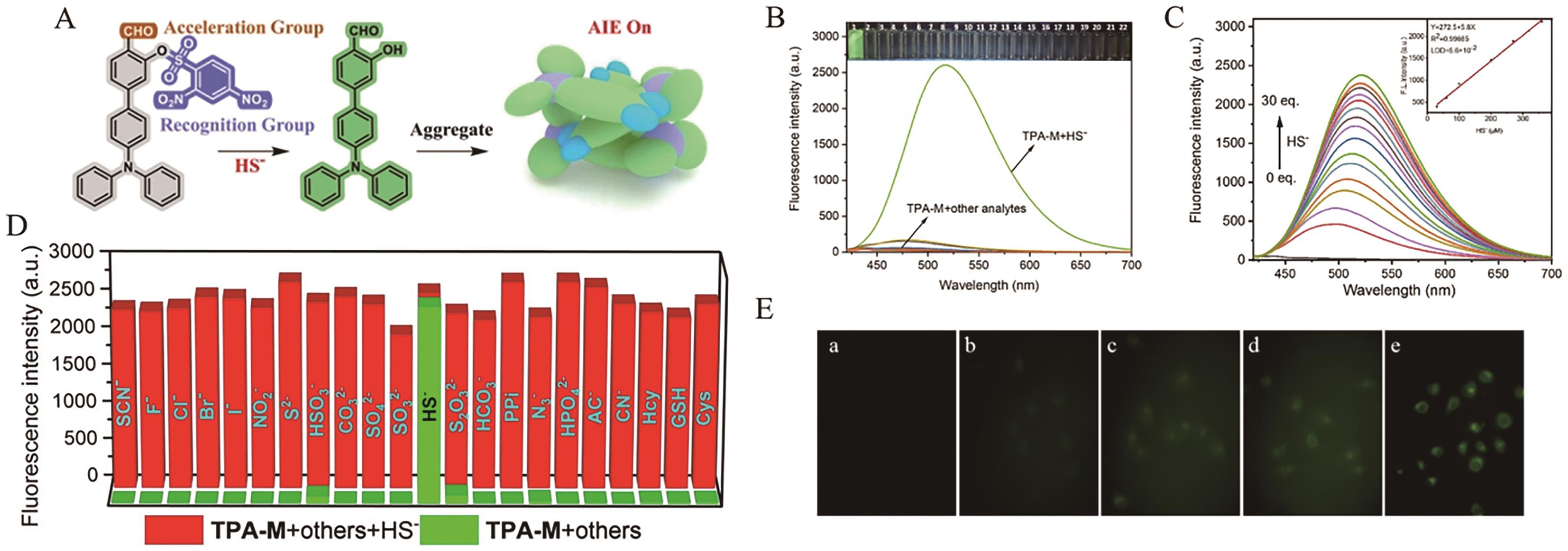
Fig.24 (A) The structure and response mechanism of AIE probe TPA-M; (B) The fluorescence response of probe TPA-M (10 μmol/L) after incubation with different analytes; (C) The fluorescence spectra of TPA-M (10 μmol/L) after reaction with various concentrations of H2S and corresponding linear relationship; (D) The fluorescence response of probe TPA-M (10 μmol/L) after incubation with different analytes in the presence of H2S(300?μmol/L); (E) The fluorescence imaging of MCF-7 cells incubated with probe TPA-M[83]
| [1] | GRIFFITHS H R, MØLLER L, BARTOSZ G, et al. Biomarkers[J]. Mol Asp Med, 2022, 23: 101-208. |
| [2] | GU X, KWOK R T K, LAM J W Y, et al. AIEgens for biological process monitoring and disease theranostics[J]. Biomaterials, 2017, 146: 115-135. |
| [3] | CULLEN N C, LEUZY A, JANELIDZE S, et al. Plasma biomarkers of Alzheimer′s disease improve prediction of cognitive decline in cognitively unimpaired elderly populations[J]. Nat Commun, 2021, 12(1): 3555-3563. |
| [4] | BROZA Y Y, ZHOU X, YUAN M, et al. Disease detection with molecular biomarkers: from chemistry of body fluids to nature-inspired chemical sensors[J]. Chem Rev, 2019, 119(22): 11761-11817. |
| [5] | KIM K, LEE C H, PARK C B. Chemical sensing platforms for detecting trace-level Alzheimer's core biomarkers[J]. Chem Soc Rev, 2020, 49(15): 5446-5472. |
| [6] | WHITFIELD M L, GEORGE L K, GRANT G D, et al. Common markers of proliferation[J]. Nat Rev Cancer, 2006, 6(2): 99-106. |
| [7] | ZHOU Y, TAO L, QIU J, et al. Tumor biomarkers for diagnosis, prognosis and targeted therapy[J]. Sig Transduct Target Ther, 2024, 9(1): 132-217. |
| [8] | WEISSLEDER R, PITTET M J. Imaging in the era of molecular oncology[J]. Nature, 2008, 452(7187): 580-589. |
| [9] | WEISSLEDER R, NAHRENDORF M. Advancing biomedical imaging[J]. Proc Natl Acad Sci, 2015, 112(47): 14424-14428. |
| [10] | HERREMANS E, MELADO-HERREROS A, DEFRAEYE T, et al. Comparison of X-ray CT and MRI of watercore disorder of different apple cultivars[J]. Postharvest Biol Tec, 2014, 87: 42-50. |
| [11] | QU H, FAN C, CHEN M, et al. Recent advances of fluorescent biosensors based on cyclic signal amplification technology in biomedical detection[J]. J Nanobiotechnol, 2021, 19(1): 403-430. |
| [12] | TANG Y, PEI F, LU X, et al. Recent advances on activatable NIR‐Ⅱ fluorescence probes for biomedical imaging[J]. Adv Opt Mater, 2019, 7(21): 1900917. |
| [13] | WANG S, LI X, CHONG S Y, et al. In vivo three‐photon imaging of lipids using ultrabright fluorogens with aggregation‐induced emission[J]. Adv Mater, 2021, 33(11): 20200749. |
| [14] | XIAO D, QI H, TENG Y, et al. Advances and challenges of fluorescent nanomaterials for synthesis and biomedical applications[J]. Nanoscale Res Lett, 2021, 16(1): 167-179. |
| [15] | XU Y, XU R, WANG Z, et al. Recent advances in luminescent materials for super-resolution imaging via stimulated emission depletion nanoscopy[J]. Chem Soc Rev, 2021, 50(1): 667-690. |
| [16] | GOPIKRISHNA P, MEHER N, IYER P K. Functional 1,8-naphthalimide AIE/AIEEgens: recent advances and prospects[J]. ACS Appl Mater Interfaces, 2017, 10(15): 12081-12111. |
| [17] | XU L, JIANG X, LIANG K, et al. Frontier luminous strategy of functional silica nanohybrids in sensing and bioimaging: from ACQ to AIE[J]. Aggregate, 2021, 3(1): e121-e149. |
| [18] | LI D, YU J. AIEgens‐functionalized inorganic‐organic hybrid materials: fabrications and applications[J]. Small, 2016, 12(47): 6478-6494. |
| [19] | BEDDARD G S, PORTER G. Concentration quenching in chlorophyll[J]. Nature, 1976, 260: 366-367. |
| [20] | LUO J, XIE Z, LAM J W Y, et al. Aggregation-induced emission of 1-methyl-1,2,3,4,5-pentaphenylsilole[J]. Chem Commun, 2001, 18: 1740-1741. |
| [21] | TU Y, ZHAO Z, LAM J W Y, et al. Mechanistic connotations of restriction of intramolecular motions (RIM)[J]. Natl Sci Rev, 2021, 8(6): nwaa260. |
| [22] | ZHANG J, ZHANG H, LAM J W Y, et al. Restriction of intramolecular motion(RIM): investigating AIE mechanism from experimental and theoretical studies[J]. Chem Res Chin Univ, 2021, 37(1): 1-15. |
| [23] | WU Q, LI Y, WANG L, et al. Aggregation-induced emission: an emerging concept in brain science[J]. Biomaterials, 2022, 286: 121581. |
| [24] | ZHAO Y Q, YU L, ZHANG L, et al. Activated aggregation‐induced emission therapeutics agents for triggering regulated cell death[J]. Aggregate, 2024, 5(3): e503-e530. |
| [25] | WANG H, LI Q, ALAM P, et al. Aggregation-induced emission (AIE), life and health[J]. ACS Nano, 2023, 17(15): 14347-14405. |
| [26] | REN K, ZHANG B, GUO J, et al. Aggregation-induced emission (AIE) for next-generation biosensing and imaging: a review[J]. Biosens Bioelectron, 2025, 271: 117067. |
| [27] | GONG S, QIN A, ZHANG Y, et al. A new ratiometric AIE fluorescent probe for detecting cysteine in food samples and imaging in the biological system[J]. Food Chem, 2023, 400: 134108. |
| [28] | ZHAO H, LI N, MA C, et al. An AIE probe for long-term plasma membrane imaging and membrane-targeted photodynamic therapy[J]. Chin Chem Lett, 2023, 34(4): 107699. |
| [29] | DUO Y, LUO G, ZHANG W, et al. Noncancerous disease-targeting AIEgens[J]. Chem Soc Rev, 2023, 52(3): 1024-1067. |
| [30] | ZHANG J, CHAI X, HE X P, et al. Fluorogenic probes for disease-relevant enzymes[J]. Chem Soc Rev, 2019, 48(2): 683-722. |
| [31] | SHARMA U, PAL D, PRASAD R. Alkaline phosphatase: an overview[J]. Ind J Clin Biochem, 2013, 29(3): 269-278. |
| [32] | SMITH B A H, BERTOZZI C R. The clinical impact of glycobiology: targeting selectins, siglecs and mammalian glycans[J]. Nat Rev Drug Discovery, 2021, 20(3): 217-243. |
| [33] | RADER B A. Alkaline phosphatase, an unconventional immune protein[J]. Front Immunol, 2017, 8: 897-902. |
| [34] | LAM K W K, CHAU J H C, YU E Y, et al. An alkaline phosphatase-responsive aggregation-induced emission photosensitizer for selective imaging and photodynamic therapy of cancer cells[J]. ACS Nano, 2023, 17(8): 7145-7156. |
| [35] | LI H, YAO Q, XU F, et al. An activatable AIEgen probe for high-fidelity monitoring of overexpressed tumor enzyme activity and its application to surgical tumor excision[J]. Angew Chem Int Ed, 2020, 59(25): 10186-10195. |
| [36] | CAI Y, ZHOU H, ZHU Y, et al. Elimination of senescent cells by β-galactosidase-targeted prodrug attenuates inflammation and restores physical function in aged mice[J]. Cell Res, 2020, 30(7): 574-589. |
| [37] | GU K, QIU W, GUO Z, et al. An enzyme-activatable probe liberating AIEgens: on-site sensing and long-term tracking of β-galactosidase in ovarian cancer cells[J]. Chem Sci, 2019, 10(2): 398-405. |
| [38] | FAN F, ZHANG L, ZHOU X, et al. A sensitive fluorescent probe for β-galactosidase activity detection and application in ovarian tumor imaging[J]. J Mater Chem B, 2021, 9(1): 170-175. |
| [39] | TIAN H, LIN W, HU XL, et al. Ratiometric sensing of β-galactosidase based on excited-state intramolecular proton transfer (ESIPT) and solid-state luminescence enhancement[J]. Org Chem Front, 2023, 10(12): 2913-2917. |
| [40] | FENG B, CHU F, HUANG X, et al. Debut of a NIR ESIPT-based fluorescent probe with synergistic effects for boosting high-contrast imaging of β-galactosidase in ovarian cancer[J]. Sens Actuators B, 2023, 396: 134541. |
| [41] | OUYANG J, SUN L, ZENG Z, et al. Nanoaggregate probe for breast cancer metastasis through multispectral optoacoustic tomography and aggregation‐induced NIR-Ⅰ/Ⅱ fluorescence imaging[J]. Angew Chem Int Ed, 2019, 59(25): 10111-10121. |
| [42] | SHEN D, DING S, LU Q, et al. Nitroreductase-responsive fluorescent “off-on” photosensitizer for hypoxic tumor imaging and dual-modal therapy[J]. ACS Omega, 2024, 9(28): 30685-30697. |
| [43] | VALLEE B L, FALCHUK K H. The biochemical basis of zinc physiology[J]. Physiol Rev, 1993, 73: 79-118. |
| [44] | CHEN X, CAI Q, LIANG R, et al. Copper homeostasis and copper-induced cell death in the pathogenesis of cardiovascular disease and therapeutic strategies[J]. Cell Death Dis, 2023, 14(2): 105-116. |
| [45] | HAMA A K H, MUSTAFA F S, OMER K M, et al. Heavy metal pollution in the aquatic environment: efficient and low-cost removal approaches to eliminate their toxicity: a review[J]. RSC Adv, 2023, 13(26): 17595-17610. |
| [46] | FREDERICKSON C J, KOH J Y, BUSH A I. The neurobiology of zinc in health and disease[J]. Nat Rev Neurosci, 2005, 6(6): 449-462. |
| [47] | XU J, XIONG J, QIN Y, et al. A novel quinolinyl-tetraphenylethene-based fluorescence “turn-on” sensor for Zn2+ with a large stokes shift and its applications for portable test strips and biological imaging[J]. Mater Chem Front, 2020, 4(11): 3338-3348. |
| [48] | JOY F, CHAITHRA K P, NIZAM A, et al. A Multi-Stimuli responsive organic luminogen with aggregation induced emission for the selective detection of Zn2+ ions in solution and solid state[J]. Chem Eng J, 2023, 453: 139798. |
| [49] | GARZA N M, SWAMINATHAN A B, MAREMANDA K P, et al. Mitochondrial copper in human genetic disorders[J]. Trends Endocrinol Metab, 2023, 34(1): 21-33. |
| [50] | RAMCHANDANI D, BERISA M, TAVAREZ D A, et al. Copper depletion modulates mitochondrial oxidative phosphorylation to impair triple negative breast cancer metastasis[J]. Nat Commun, 2021, 12(1): 7311-7326. |
| [51] | CAI X M, LI S, WANG W J, et al. Natural acceptor of coumarin-isomerized red-emissive bioAIEgen for monitoring Cu2+ concentration in live cells via FLIM[J]. Adv Sci, 2023, 11(9): 2307078. |
| [52] | JIANG J, SUN H, HU Y, et al. AIE+ESIPT activity-based NIR Cu2+ sensor with dye participated binding strategy[J]. Chem Commun, 2021, 57(62): 7685-7688. |
| [53] | NIU X, ZHANG H, WU X, et al. An AIE-active “turn-off” fluorescent sensor for highly selective and sensitive detection of Cu2+ ions[J]. J Mol Struct, 2022, 1264: 133294. |
| [54] | NESCI S, TROMBETTI F, PIRINI M, et al. Mercury and protein thiols: stimulation of mitochondrial F1FO-ATPase and inhibition of respiration[J]. Chem Biol Interact, 2016, 260: 42-49. |
| [55] | KIRAN, BHARTI R, SHARMA R. Effect of heavy metals: an overview[J]. Mater Today: Proc, 2022, 51: 880-885. |
| [56] | TONAZZI A, GIANGREGORIO N, CONSOLE L, et al. Mitochondrial carnitine/acylcarnitine transporter, a novel target of mercury toxicity[J]. Chem Res Toxicol, 2015, 28(5): 1015-1022. |
| [57] | TAN T, ZHANG C, HAN Y, et al. Fine-tuning bromide AIE probes for Hg2+ detection in mitochondria with wash-free staining[J]. J Hazard Mater, 2024, 464: 132999. |
| [58] | WU S, YANG Y, CHENG Y, et al. Fluorogenic detection of mercury ion in aqueous environment using hydrogel-based AIE sensing films[J]. Aggregate, 2022, 4(3): e287-e294. |
| [59] | WALLACE D C. Mitochondrial diseases in man and mouse[J]. Science, 1999, 283: 1482-1488. |
| [60] | YANG B, CHEN Y, SHI J. Reactive oxygen species (ROS)-based nanomedicine[J]. Chem Rev, 2019, 119(8): 4881-4985. |
| [61] | GRIFFITHS H R, GAO D, PARARASA C. Redox regulation in metabolic programming and inflammation[J]. Redox Biol, 2017, 12: 50-57. |
| [62] | BEGHETTO C, RENKEN C, ERIKSSON O, et al. Implications of the generation of reactive oxygen species by photoactivated calcein for mitochondrial studies[J]. Eur J Biochem, 2000, 267: 5585-5592 |
| [63] | RYTER S W, TYRRELL R M. Singlet molecular oxygen (1O2) a possible effector of eukaryotic gene expression[J]. Free Radic Biol Med, 1998, 24: 1520-1534. |
| [64] | KLOTZ L O, BRIVIBA K, SIES H. Mitogen-activated protein kinase activation by singlet oxygen and ultraviolet A[J]. Methods Enzymol, 2000, 319: 130-143. |
| [65] | LIU C, WANG X, LIU J, et al. Near‐infrared AIE dots with chemiluminescence for deep-tissue imaging[J]. Adv Mater, 2020, 32(43): 2004685. |
| [66] | LYU J, CHENG M, LIU J, et al. An aggregation-induced emission nanosensor for real-time chemiluminescent sensing of light-independent intracellular singlet oxygen[J]. ACS Appl Mater Interfaces, 2022, 14(48): 54081-54089. |
| [67] | RHEE S G. H2O2, a necessary evil for cell signaling[J]. Science, 2006, 312: 1882-1883. |
| [68] | FINKEL T, HOLBROOK N J. Oxidants, oxidative stress and the biology of ageing[J]. Nature, 2000, 408: 239-247. |
| [69] | BHOSLE A A, BANERJEE M, GUPTA V, et al. Mechanochemical synthesis of an AIE-TICT-ESIPT active orange-emissive chemodosimeter for selective detection of hydrogen peroxide in aqueous media and living cells, and solid-phase quantitation using a smartphone[J]. New J Chem, 2022, 46(39): 18961-18972. |
| [70] | CHEN J, CHEN L, WU Y, et al. A H2O2-activatable nanoprobe for diagnosing interstitial cystitis and liver ischemia-reperfusion injury via multispectral optoacoustic tomography and NIR-Ⅱ fluorescent imaging[J]. Nat Commun, 2021, 12(1): 6870-6884. |
| [71] | IWAKIRI Y, KIM M Y. Nitric oxide in liver diseases[J]. Trends Pharmacol Sci, 2015, 36(8): 524-536. |
| [72] | JIN G, GAO Z, LIU Y, et al. Polymeric nitric oxide delivery nanoplatforms for treating cancer, cardiovascular diseases, and infection[J]. Adv Healthcare Mater, 2020, 10(3): 2001550. |
| [73] | SUN L, OUYANG J, MA Y, et al. An activatable probe with aggregation‐induced emission for detecting and imaging herbal medicine induced liver injury with optoacoustic imaging and NIR‐II fluorescence imaging[J]. Adv Healthcare Mater, 2021, 10(24): 2100867. |
| [74] | CHEN J, CHEN L, FANG Y, et al. Refashioning benzothiadiazole dye as an activatable nanoprobe for biomarker detection with NIR-Ⅱ fluorescence/optoacoustic imaging[J]. Cell Reports Phys Sci, 2022, 3(2): 100570. |
| [75] | FILOMENI G, ROTILIO G, CIRIOLO M R. Cell signalling and the glutathione redox system[J]. Biochem Pharmacol, 2002, 64(5/6): 1057-1064. |
| [76] | TOWNSEND D M, TEW K D, TAPIERO H. The importance of glutathione in human disease[J]. Biomed Pharmacother, 2003, 57(3/4): 145-155. |
| [77] | DAI F, ZHAO M, YANG F, et al. An ESIPT coupled AIE fluorescent probe for biothiols detection and imaging based on a chalcone fluorophore[J]. Dyes Pigm, 2020, 183: 108627. |
| [78] | ZHANG Y H, LI X, HUANG L, et al. AIE based GSH activatable photosensitizer for imaging-guided photodynamic therapy[J]. Chem Commun, 2020, 56(71): 10317-10320. |
| [79] | SHEN Y, WEI Y, GAO X, et al. Engineering an enzymatic cascade catalytic smartphone-based sensor for onsite visual ratiometric fluorescence-colorimetric dual-mode detection of methyl mercaptan[J]. Environ Sci Technol, 2023, 57(4): 1680-1691. |
| [80] | LEE M, SCHWAB C, YU S, et al. Astrocytes produce the antiinflammatory and neuroprotective agent hydrogen sulfide[J]. Neurobiol Aging, 2009, 30(10): 1523-1534. |
| [81] | YANG G, WU L, JIANG B, et al. H2S as a physiologic vasorelaxant hypertension in mice with deletion of cystathionine γ-lyase[J]. Science, 2008, 322: 587-590. |
| [82] | GU B, DAI C, ZHOU Z, et al. Rational construction of an AIE-active fluorescent probe bearing three reaction sites for individual and continuous detection of H2S and HClO with single-wavelength excitation[J]. Sens Actuators, B, 2023, 375: 132900-132907. |
| [83] | MENG Y, YAO X, ZHONG K, et al. An aggregation-induced emission‐based fluorescence turn-on probe for efficient detection of HS- in water, wine, and living cells[J]. Eur J Org Chem, 2023, 26(18): e202300022. |
| [1] | Zhen-Zhen MENG, Jin-Feng YANG, Yu-Kang YAN. Application of Near Infrared Aggregation-Induced Emission Fluorescence Probe with Benzopyranitrile in Viscosity Detection and Cell Imaging [J]. Chinese Journal of Applied Chemistry, 2025, 42(7): 971-981. |
| [2] | Zhen-Cao WANG, Ning LYU, Liang-Fang CHEN, Yue-Feng RAO. Research Progress of Aggregation-Induced Emission Molecules for Fluorescence Imaging Therapy [J]. Chinese Journal of Applied Chemistry, 2025, 42(6): 741-756. |
| [3] | Ju-Ying XIAO, Xia ZHAO, Yuan LIN, Zhao-Hui SU. Preparation and Investigation of Functional PEGylated Chitosan Nanoparticles [J]. Chinese Journal of Applied Chemistry, 2025, 42(3): 386-395. |
| [4] | Li-Zhen YUAN, Ni-Ya LIN, Yun-Fan ZHANG, Jing-Jing HU, Xiao-Ding LOU, Fan XIA. Development in Probes on Outer Surface of Nanochannels for Detecting Biomarkers [J]. Chinese Journal of Applied Chemistry, 2024, 41(1): 87-99. |
| [5] | Da-Wei TONG, Ming KONG, Yu-Bin XIANG. Synthesis, Photophysical Properties, Theoretical Calculation and Cell Imaging of a Tetraphenylethene Imidazole Compound with Methoxy Group [J]. Chinese Journal of Applied Chemistry, 2023, 40(9): 1322-1329. |
| [6] | Yue YANG, Shi-Wen HUANG, Yue TONG, Ze-Da CHEN, Ben-Hua MA, Chuan-Dong DOU. Donor-Acceptor Type Chiral Tetracoordinate Organoboranes and Their Optical Properties [J]. Chinese Journal of Applied Chemistry, 2023, 40(5): 743-748. |
| [7] | Jia-Mei GENG, Su-Fang MA, Wen LIU, Hai-Peng DIAO, Zhi-Fang WU, Si-Jin LI. Liver-Targeted Fluorescent Probes for Specific Detection of ONOO- in HepG2 Cells [J]. Chinese Journal of Applied Chemistry, 2023, 40(3): 441-448. |
| [8] | ZHANG Nan, LI Tie, YANG Guang, HUANG Xin, YUE Hao, WANG Yang, LIU Jun-Tong, LIU Shu-Ying, WANG Fu-Chun. Screening for Urine Metabolic Biomarkers of Transient Ischemic Attack [J]. Chinese Journal of Applied Chemistry, 2021, 38(3): 305-314. |
| [9] | CHENG Jinhua, JIANG Hongji. Study on Self-assembly Behaviors of an Amphiphilic Block Polymer by Terminally Grafting Tetraphenylethene-Based Aggregation-Induced Emission Active Moietys [J]. Chinese Journal of Applied Chemistry, 2019, 36(4): 440-450. |
| [10] | WANG Tao,MA Lamaocao,MA Hengchang. Research Progress on Cell Imaging Based on the Aggregation-induced Emission Fluorescent Probes [J]. Chinese Journal of Applied Chemistry, 2018, 35(10): 1155-1165. |
| [11] | YAO Qianfang,CHENG Wenyu,YIN Meizhen. Host-Guest Supramolecular Fluorescent Probes Based on Macrocyclic Molecules [J]. Chinese Journal of Applied Chemistry, 2017, 34(12): 1344-1354. |
| [12] | BU Lulu,WANG Qing,XIE Yongshu. Research Progress of Fluorescent Zinc Probes [J]. Chinese Journal of Applied Chemistry, 2017, 34(12): 1355-1369. |
| [13] | CHEN Jianhua,LI Dongyang,LIU Shenghua,TAN Ying,YIN Jun. Recent Advances in Phosgene and Nerve Agents Responsive Fluorescent Probes [J]. Chinese Journal of Applied Chemistry, 2017, 34(12): 1413-1432. |
| [14] | Jia ZHOU, Yun NI, Chengwu ZHANG, Xinghan QIU, Yanfei ZHAO, Lei BAI, Gaobin ZHANG, Lin LI. Design and Synthesis of Pyrimidine Based Two-Photon Fluorescence Probe and Its Application in Bioimaging [J]. Chinese Journal of Applied Chemistry, 2017, 34(12): 1450-1456. |
| [15] | Jia ZHOU, Yun NI, Chengwu ZHANG, Xinghan QIU, Yanfei ZHAO, Lei BAI, Gaobin ZHANG, Lin LI. Design and Synthesis of Pyrimidine Based Two-Photon Fluorescence Probe and Its Application in Bioimaging [J]. Chinese Journal of Applied Chemistry, 2017, 34(12): 0-0. |
| Viewed | ||||||
|
Full text |
|
|||||
|
Abstract |
|
|||||