

MENG Yating, JIAO Yuan, ZHANG Yuan, et al. Synthesis of Red Emission Fluorescent Carbon Dots and Its Application for Detection of Persulfate[J]. Chinese Journal of Applied Chemistry, 37(6): 719-725


以对苯二胺为碳源,乙醇为溶剂,采用溶剂热法合成红色荧光碳点(R-CDs)。 通过透射电子显微镜、紫外可见吸收光谱(UV-Vis)、红外光谱(FT-IR)、X射线光电子能谱(XPS)对其进行表征。 合成的R-CDs粒径约为(3.63±0.20) nm。 R-CDs的最大激发和发射波长分别为480和620 nm,具有激发波长独立性,光稳定性好。 基于静态猝灭,R-CDs的荧光可以被过硫酸根(S2O82-)有效猝灭。 在2.5~120 μmol/L范围内,S2O82-的浓度与R-CDs的荧光猝灭程度呈线性关系,相关系数( R2)为0.9970,检出限为1.2 μmol/L,具有良好的选择性和高灵敏度。 同时,该荧光传感体系可应用于自来水和湖水样品中S2O82-的检测。
Red fluorescent carbon dots (R-CDs) were synthesized by one-step solvothermal method by using p-phenylenediamine and ethanol. The synthesized R-CDs were characterized by transmission electron microscopy (TEM), ultraviolet-visible spectroscopy (UV-Vis), Fourier transform infrared spectroscopy (FTIR), X-ray photoelectron spectroscopy (XPS) and fluorescence spectroscopy, respectively. The results indicate that the R-CDs are uniform with an average size of (3.63±0.20) nm. Abundant groups like hydroxyl and amine groups are linked on the surface of the synthesized R-CDs. The as-prepared R-CDs show excitation-independent property and the maximum excitation and emission wavelengths are 480 and 620 nm, respectively. Based on static quenching between R-CDs and persulfate, the fluorescence of R-CDs could be effectively quenched by persulfate. A fluorescent strategy is developed for detection of persulfate. The method showed a linear range of 2.5~120 μmol/L with a correlation coefficient ( R2) of 0.9970. The limit of detection was 1.2 μmol/L, showing excellent sensitivity and selectivity. Meanwhile, the as-proposed sensing system is successfully applied to the analysis of persulfate in tap water and lake water samples with satisfactory results.


过硫酸盐是一种强氧化剂,已应用于水和土壤去污剂[1]、电路板制作[2]、化妆品[3]和聚合[4]等领域。 据报道,过硫酸盐可能导致或加剧哮喘[5]和皮肤过敏[6]等疾病,因此对过硫酸盐进行检测具有重要意义。 目前,测定过硫酸盐的方法限于电化学法[7,8,9],因其存在操作繁琐,响应时间较长等缺陷,从而限制了该方法的应用。 由于荧光传感方法具有灵敏度高、选择性好和易于操作等优势,因此发展过硫酸盐的荧光检测方法十分必要。
碳点(CDs)作为一种新型碳纳米材料,由于其独特的光学性质[10]、优异的水溶性[11]、低毒性[12]和良好的生物相容性[13]被广泛应用于生物成像[14]、分析检测[15]和光电器件[16]等领域。 长波长发射的碳点由于其能够克服短波背景干扰的优点引起了广泛关注,构筑红色荧光发射的碳点是当前碳点发展的趋势[17]。 目前,一些文献报道了长波长发射碳点的设计策略及相关荧光传感,实现了金属阳离子[18],氨基酸[19]以及生物小分子[20]的灵敏检测。 据我们所知,碳点用于检测过硫酸根阴离子的研究还未见报道。
本文在文献[21]的基础上,以对苯二胺为前驱体,乙醇为溶剂,采用一步溶剂热法合成了具有激发波长独立的红色荧光碳点(R-CDs)。 所制备的R-CDs具有良好的水溶性、光稳定性。 基于静态猝灭原理,R-CDs的荧光可以被S2O82-有效猝灭。 据此,我们建立了基于碳点的S2O82-荧光检测方法,并将其进一步应用于实际样品中S2O82-的检测。
JEM-2100型透射电子显微镜(TEM,日本电子株式会社); Bruker Tensor Ⅱ型傅里叶红外光谱仪(FTIR,德国 Bremen 公司);Axis Ultra Dld X型射线光电子能谱(XPS,英国 Axis Ultradld 公司); F-4500型荧光分光光度计(PS,日本日立公司); UV-265型紫外-可见分光光度计(UV-Vis,日本岛津公司); FLS920 型爱丁堡全功能型稳态瞬态荧光光谱仪(英国 Edinburgh Instruments公司);pH计(瑞士梅特勒-托利多公司)。
对苯二胺(97%)、过硫酸钾(99%)、罗丹明B(95%)购自阿拉丁试剂有限公司;其它试剂均为国产分析纯;实验用水均是MilliQ Plus系统制备的超纯水(≥18.25 MΩ·cm)。
1.2.1 R-CDs的合成方法
将0.2 g对苯二胺溶解于20 mL乙醇中,在磁力搅拌下搅拌20 min,超声得到澄清溶液。 然后,将其转移至50 mL水热反应釜中,在200 ℃下加热6 h。 过滤不溶物后得到红色溶液。 通过截留相对分子质量500~1000的透析袋,透析处理3 d,得到纯净的碳点水溶液。 将上述碳点水溶液冷冻干燥后得到碳点粉末。
1.2.2 荧光量子产率的测定
以罗丹明B的乙醇溶液(量子产率QY=0.89)为参比物通过下式计算R-CDs的荧光量子产率:
式中,R为罗丹明B的相关参数, Φ 为荧光量子产率(%), n为积分荧光强度, A为入射光吸光度, η为溶剂的折射率。
1.2.3 S2O82-的荧光检测及选择性
在1 cm石英比色皿中依次加入2 mL R-CDs水溶液(0.25 mg/mL),10 μL不同浓度的S2O82-,测量其发射光谱。 以其它阴离子代替S2O82-,研究R-CDs对S2O82-的选择性。
1.2.4 实际样品中S2O82-的的检测
实际样品为山西大学校内自来水和令德湖水,水样隔夜静置,经0.22 μm 滤膜过滤后,采用本方法测样品中S2O82-的含量。 在样品中分别加入不同浓度的S2O82-,测定加标回收率。
TEM表征结果如图1A所示,R-CDs呈准球形,均匀分散,平均粒径约为(3.63±0.20) nm。 红外光谱(图1B)显示,R-CDs中存在以下官能团:OH/NH(3681和3292 cm-1)、CH(2865和2945 cm-1)、C—NH(1360~1457 cm-1)和C—OH(1031和1078 cm-1)。 R-CDs的光电子能谱(XPS)如图1C所示,在285、400和231 eV处的特征峰分别对应C1 s、N1 s和O1 s。 R-CDs的C1 s XPS光谱包含2个峰:284.7 eV(C=C)和286.0 eV(C—O)(图1D)。 N1 s XPS光谱中399.1和400.8 eV处的峰分别对应N—H和C—N—C基团(图1E)。 O1 s XPS 光谱可以分解为2个峰:531.8和533.3 eV,分别对应C=O和C—O—C基团(图1F)。 以上结果表明,R-CDs表面存在氨基、羟基官能团。
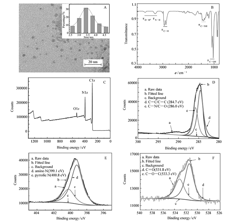 | 图1 R-CDs的TEM图(A,插图为粒径统计分布),红外光谱(B)和X射线光电子能谱全谱(C),高分辨率C1 s(D)、N1 s(E)和O1 s X射线光电子能谱(F)Fig.1 TEM image (A, inset is the histogram of particle size distribution), FTIR(B), XPS survey scan(C), C1 s(D), N1 s(E), O1 s(F) spectra of R-CDs |
由紫外-可见吸收光谱(图 2A)可见,R-CDs溶液在258和285 nm处有明显吸收峰, 归因于芳香族C=C双键发生的 π-π*跃迁,并表明芳香杂环的存在。 在496 nm处的吸收峰可能归因于R-CDs表面官能团的 n-π*跃迁。 R-CDs的最大激发和发射波长为480和620 nm。 如图2B 所示,当激发波长从300 nm 增加至600 nm,发射峰未发生红移,显示出激发波长独立性。
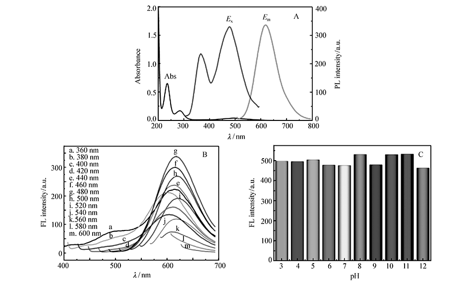 | 图2 R-CDs的紫外-可见吸收光谱、最大激发和发射光谱(A);不同激发波长下的发射光谱(B);pH值对R-CDs荧光强度的影响(C)Fig.2 (A)UV-Vis absorption and photoluminescence excitation and emission spectra of R-CDs. (B)Fluorescence emission spectra of R-CDs at different excitation wavelengths. (C)Effect of pH on the fluorescence intensity of R-CDs aqueous |
考察了溶液pH值和氙灯照射时间对R-CDs稳定性的影响。 如图2C所示,在pH值3~11的范围内,R-CDs的荧光强度几乎不变。 用氙灯连续照射60 min,R-CDs的荧光强度未发生明显改变,表现出良好的光稳定性。 以罗丹明B为参比物,计算R-CDs在激发波长为480 nm的量子产率(QY)8.8%。
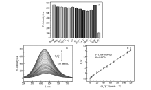
分析了12种阴离子对R-CDs荧光强度的影响。 如图3A所示,加入S2O82-后,R-CDs的荧光强度明显降低。 而其它阴离子的加入对R-CDs的荧光强度几乎无影响,表明R-CDs对S2O82-有良好的选择性。 如图3B所示,随着S2O82-浓度的增加,R-CDs的荧光强度逐渐降低。 S2O82-浓度在2.5~120 μmol/L范围内与R-CDs的荧光猝灭程度呈线性关系,线性回归方程为 y=0.0042 x+1.014,相关系数( R2)为0.9970,检出限为1.2 μmol/L。 不同方法对S2O82-的检测性能对比见表1,表中其它方法均为电化学检测法。 基于R-CDs,首先建立S2O82-的荧光检测方法。
| 表1 不同方法对S2O82-检测性能的比较 Table 1 Comparison of S2O82- detection performance of different methods |
 | 图3 (A)R-CDs对S2O82-的选择性;(B)不同浓度的S2O82-对R-CDs荧光光谱的影响;(C) F0 /F和S2O82-浓度之间的线性关系Fig.3 (A)Effect of different ions on fluorescence intensity of R-CDs. (B)The quenching of photoluminescence of R-CDs in the presence of S2O82-. (C)The linear correlation of F0 /F values versus S2O82- concentration |
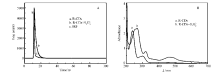
为了探究S2O82-对R-CDs的猝灭过程,分别测定了加入S2O82-前后R-CDs的荧光寿命。 通过双指数函数拟合荧光衰减曲线,如图4A所示。 R-CDs的平均荧光寿命为2.52 ns,加入S2O82-后,平均荧光寿命为2.92 ns,无明显变化。 随着S2O82-的加入,R-CDs的紫外可见吸收光谱发生改变。 如图4B所示,R-CDs在255、269、320和470 nm处出现新的吸收峰,证明S2O82-和R-CDs结合形成基态配合物。 此外,由图2C可以得出,Stern-Volmer动态猝灭常数 Ksv为0.0042 L/mol。 由式(2)可以得到 Kq=1.68×10¹² L/(mol·s),远大于2.0×1010 L/(mol·s)。 因此,推断S2O82-对R-CDs的猝灭过程为静态猝灭。
式中, F0表示未加入猝灭剂S2O82-时的荧光强度, F表示加入猝灭剂S2O82-后的荧光强度, cQ表示猝灭剂S2O82-的浓度(mol/L), τ0表示未加入猝灭剂S2O82-时碳点的平均荧光寿命(ns), Kq表示双分子猝灭过程速率常数(L/(mol·s)), Ksv是Stern-Volmer动态猝灭常数(L/mol)。
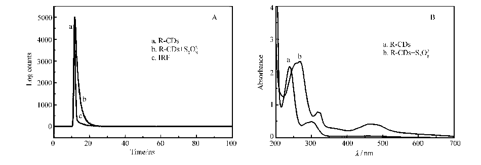 | 图4 加入S2O82-前后R-CDs的荧光寿命衰减曲线(A)和紫外吸收光谱(B)Fig.4 (A)Fluorescence decay and (B)UV-Vis absorption spectra of R-CDs in the absence and presence of S2O82- |
取校内自来水和令德湖水,按1.2.4节中的方法处理后,采用本方法测样品中S2O82-的含量,结果见表2,回收率在97.8%~104.0%,相对标准偏差(RSD)在1.9%~3.5%,表明该方法可以用于实际样品中S2O82-的检测。
| 表2 水样中S2O82-的检测结果 Table 2 The determination results of S2O82- in water samples |
通过一步溶剂热法合成激发波长独立的红色荧光碳点(R-CDs),所制备的碳点具有良好的水溶性和优异的光稳定性。 基于静态猝灭,R-CDs的荧光可以被S2O82-有效猝灭,首先建立基于碳点的S2O82-荧光检测方法。 该方法选择性好、灵敏度高,并进一步用于自来水和令德湖水样品中S2O82-的检测。
| [1] |
|
| [2] |
|
| [3] |
|
| [4] |
|
| [5] |
|
| [6] |
|
| [7] |
|
| [8] |
|
| [9] |
|
| [10] |
|
| [11] |
|
| [12] |
|
| [13] |
|
| [14] |
|
| [15] |
|
| [16] |
|
| [17] |
|
| [18] |
|
| [19] |
|
| [20] |
|
| [21] |
|